Introduction – Company Background
GuangXin Industrial Co., Ltd. is a specialized manufacturer dedicated to the development and production of high-quality insoles.
With a strong foundation in material science and footwear ergonomics, we serve as a trusted partner for global brands seeking reliable insole solutions that combine comfort, functionality, and design.
With years of experience in insole production and OEM/ODM services, GuangXin has successfully supported a wide range of clients across various industries—including sportswear, health & wellness, orthopedic care, and daily footwear.
From initial prototyping to mass production, we provide comprehensive support tailored to each client’s market and application needs.
At GuangXin, we are committed to quality, innovation, and sustainable development. Every insole we produce reflects our dedication to precision craftsmanship, forward-thinking design, and ESG-driven practices.
By integrating eco-friendly materials, clean production processes, and responsible sourcing, we help our partners meet both market demand and environmental goals.
Core Strengths in Insole Manufacturing
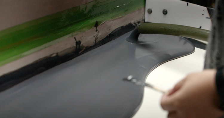
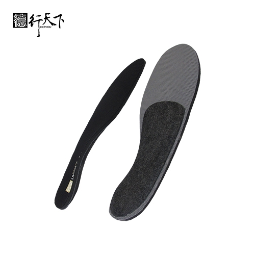
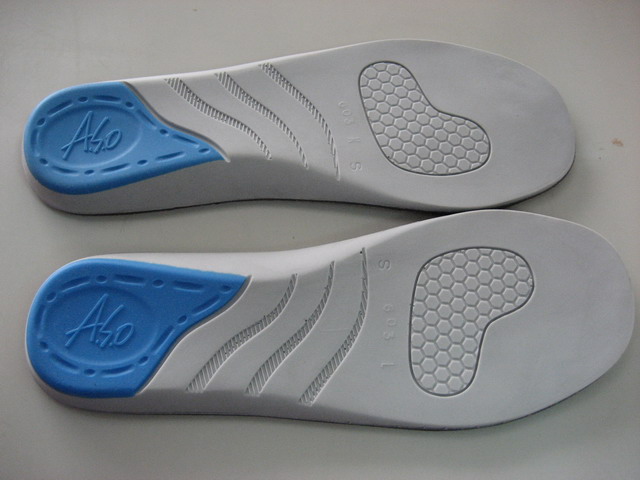
At GuangXin Industrial, our core strength lies in our deep expertise and versatility in insole and pillow manufacturing. We specialize in working with a wide range of materials, including PU (polyurethane), natural latex, and advanced graphene composites, to develop insoles and pillows that meet diverse performance, comfort, and health-support needs.
Whether it's cushioning, support, breathability, or antibacterial function, we tailor material selection to the exact requirements of each project-whether for foot wellness or ergonomic sleep products.
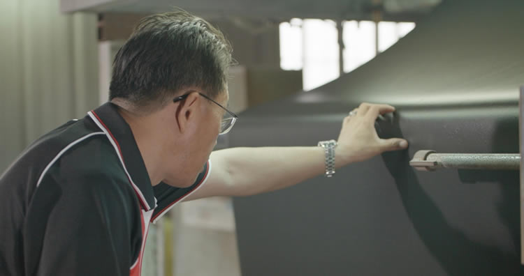
We provide end-to-end manufacturing capabilities under one roof—covering every stage from material sourcing and foaming, to precision molding, lamination, cutting, sewing, and strict quality control. This full-process control not only ensures product consistency and durability, but also allows for faster lead times and better customization flexibility.
With our flexible production capacity, we accommodate both small batch custom orders and high-volume mass production with equal efficiency. Whether you're a startup launching your first insole or pillow line, or a global brand scaling up to meet market demand, GuangXin is equipped to deliver reliable OEM/ODM solutions that grow with your business.
Customization & OEM/ODM Flexibility
GuangXin offers exceptional flexibility in customization and OEM/ODM services, empowering our partners to create insole products that truly align with their brand identity and target market. We develop insoles tailored to specific foot shapes, end-user needs, and regional market preferences, ensuring optimal fit and functionality.
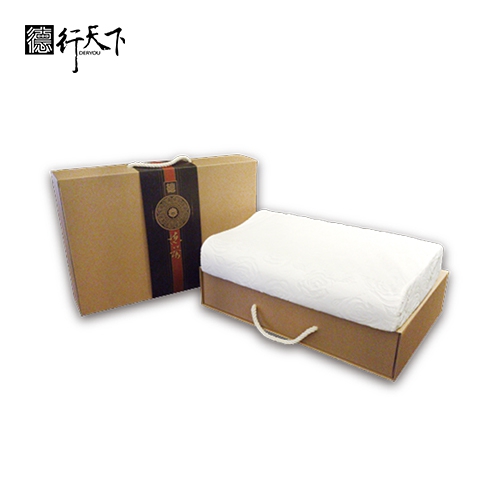
Our team supports comprehensive branding solutions, including logo printing, custom packaging, and product integration support for marketing campaigns. Whether you're launching a new product line or upgrading an existing one, we help your vision come to life with attention to detail and consistent brand presentation.
With fast prototyping services and efficient lead times, GuangXin helps reduce your time-to-market and respond quickly to evolving trends or seasonal demands. From concept to final production, we offer agile support that keeps you ahead of the competition.
Quality Assurance & Certifications
Quality is at the heart of everything we do. GuangXin implements a rigorous quality control system at every stage of production—ensuring that each insole meets the highest standards of consistency, comfort, and durability.
We provide a variety of in-house and third-party testing options, including antibacterial performance, odor control, durability testing, and eco-safety verification, to meet the specific needs of our clients and markets.
Our products are fully compliant with international safety and environmental standards, such as REACH, RoHS, and other applicable export regulations. This ensures seamless entry into global markets while supporting your ESG and product safety commitments.
ESG-Oriented Sustainable Production
At GuangXin Industrial, we are committed to integrating ESG (Environmental, Social, and Governance) values into every step of our manufacturing process. We actively pursue eco-conscious practices by utilizing eco-friendly materials and adopting low-carbon production methods to reduce environmental impact.
To support circular economy goals, we offer recycled and upcycled material options, including innovative applications such as recycled glass and repurposed LCD panel glass. These materials are processed using advanced techniques to retain performance while reducing waste—contributing to a more sustainable supply chain.
We also work closely with our partners to support their ESG compliance and sustainability reporting needs, providing documentation, traceability, and material data upon request. Whether you're aiming to meet corporate sustainability targets or align with global green regulations, GuangXin is your trusted manufacturing ally in building a better, greener future.
Let’s Build Your Next Insole Success Together
Looking for a reliable insole manufacturing partner that understands customization, quality, and flexibility? GuangXin Industrial Co., Ltd. specializes in high-performance insole production, offering tailored solutions for brands across the globe. Whether you're launching a new insole collection or expanding your existing product line, we provide OEM/ODM services built around your unique design and performance goals.
From small-batch custom orders to full-scale mass production, our flexible insole manufacturing capabilities adapt to your business needs. With expertise in PU, latex, and graphene insole materials, we turn ideas into functional, comfortable, and market-ready insoles that deliver value.
Contact us today to discuss your next insole project. Let GuangXin help you create custom insoles that stand out, perform better, and reflect your brand’s commitment to comfort, quality, and sustainability.
🔗 Learn more or get in touch:
🌐 Website: https://www.deryou-tw.com/
📧 Email: shela.a9119@msa.hinet.net
📘 Facebook: facebook.com/deryou.tw
📷 Instagram: instagram.com/deryou.tw
Graphene insole OEM factory Vietnam
Are you looking for a trusted and experienced manufacturing partner that can bring your comfort-focused product ideas to life? GuangXin Industrial Co., Ltd. is your ideal OEM/ODM supplier, specializing in insole production, pillow manufacturing, and advanced graphene product design.
With decades of experience in insole OEM/ODM, we provide full-service manufacturing—from PU and latex to cutting-edge graphene-infused insoles—customized to meet your performance, support, and breathability requirements. Our production process is vertically integrated, covering everything from material sourcing and foaming to molding, cutting, and strict quality control.ESG-compliant OEM manufacturer in Thailand
Beyond insoles, GuangXin also offers pillow OEM/ODM services with a focus on ergonomic comfort and functional innovation. Whether you need memory foam, latex, or smart material integration for neck and sleep support, we deliver tailor-made solutions that reflect your brand’s values.
We are especially proud to lead the way in ESG-driven insole development. Through the use of recycled materials—such as repurposed LCD glass—and low-carbon production processes, we help our partners meet sustainability goals without compromising product quality. Our ESG insole solutions are designed not only for comfort but also for compliance with global environmental standards.Insole ODM factory in Taiwan
At GuangXin, we don’t just manufacture products—we create long-term value for your brand. Whether you're developing your first product line or scaling up globally, our flexible production capabilities and collaborative approach will help you go further, faster.China graphene sports insole ODM
📩 Contact us today to learn how our insole OEM, pillow ODM, and graphene product design services can elevate your product offering—while aligning with the sustainability expectations of modern consumers.Indonesia OEM factory for footwear and bedding
Recent research in the Arctic reveals that jellyfish, once considered negligible in food webs, are a key food source for amphipods during the polar night in Svalbard’s Kongsfjorden, indicating significant changes in the ecosystem due to “Atlantification.” Pictures is a ctenophore or comb jelly. Credit: C. Havermans AWI research team shows that jellyfish play an important, previously unknown role in the diet of amphipods during the polar night. The Arctic is changing rapidly due to climate change. It is not only affected by increasing surface temperatures, but also by warm water from the Atlantic, which is flowing in more and more – changing the structures and functions of the ecosystem as it also leads to species from warmer regions, such as jellyfish, arriving in the Arctic. Using DNA metabarcoding, researchers from the Alfred Wegener Institute have now been able to prove for the first time that these jellyfish serve as food for amphipods on Svalbard during the polar night and thus play a greater role in Arctic food webs than previously assumed. They present their findings in a recent article in the scientific journal Frontiers in Marine Science. The AWI researchers collected samples from four different amphipod species over the course of a month during the polar night. Credit: Alfred Wegener Institute / Charlotte Havermans Atlantification of the Arctic and Its Impact on Marine Life In recent years, warm, salty water from the Atlantic has increasingly found its way into the European Arctic. The Norwegian archipelago of Svalbard is also under the influence of this “Atlantification”: the Kongsfjorden on the west coast has switched to an Atlantic regime; the water temperature during the polar night (November to February) is increasing by around 2 degrees Celsius per decade. These changes also lead to biotic shifts, as species from warmer waters also flow into the Arctic along with the warm Atlantic water. “Some jellyfish species in particular tend to spread poleward and into the Arctic,” says Charlotte Havermans, head of the ARJEL junior research group at the Alfred Wegener Institute, Helmholtz Center for Polar and Marine Research (AWI). ” When we were in Kongsfjorden in the Polar Night in 2022, we were very surprised to see the fjord teeming with jellyfish life, consisting of many different species and life stages, and they seemed to be the dominant zooplankton in winter time.” Pink helmet jellyfish, a hydrozoan. Credit: C. Havermans Jellyfish in Arctic Food Webs In the past, jellyfish were considered a trophic dead end in marine food webs, but recent studies suggest that they are an important prey for marine invertebrates and fish. “Therefore, we wondered whether the jellyfish in Kongsfjorden also serve as food for other organisms, especially during the dark season of the polar night when other food sources are limited,” says Havermans. To answer this question, one of the team’s PhD students, Annkathrin Dischereit, analyzed the stomach contents of various amphipod species. For a month, they regularly collected samples from four different amphipod species (Gammarus oceanicus, G. setosus, Orchomenella minuta, and Anonyx sarsi) during the polar night, using baited traps and hand nets. Jellyfish Are an Integral Part of the Diet of Amphipods During the Polar Night The AWI researchers used DNA-metabarcoding to determine the food spectrum of the small crustaceans. This cutting-edge method can detect short gene fragments in the stomach, which are then compared with genetic reference databases to identify the prey species to which the fragments belong. “We found a large number of jellyfish in the stomachs of the amphipods, from the largest jellyfish in the fjord to tiny hydrozoans,” explains Charlotte Havermans. Using DNA metabarcoding, the AWI team was able to identify and categorize the soft parts of jellyfish and other organisms that had been consumed, even if they were already heavily digested. “We were able to prove for the first time that amphipod scavengers feed on the remains of jellyfish. This had previously only been shown in experimental environments.” All the species studied fed on both plant and animal matter. In addition to jellyfish, crustaceans, and macroalgae were other important components of the diet of some species, while fish species such as the polar cod or snailfish played an important role for other species. Whether the amphipods fed on eggs, larvae, carrion, or feces of fish remains to be clarified. What also remains to be determined, is whether jellyfish act as a survival food in winter, or are part of the regular prey of these organisms in all seasons. “We have always assumed that the nutritional value of jellyfish is low, but this has only been investigated for less than a handful of species, and also depends on the tissues that are utilized.” New Insights Into Arctic Marine Ecosystems The study provides completely new insights into the Arctic food web during the polar night and are the first natural, non-experimental evidence for the role of jellyfish in these webs. “The thriving, diverse jellyfish community that occurs in Kongsfjorden in winter is clearly used as a food source,” Charlotte Havermans summarises the results. “Until now, we knew nothing about the role of jellyfish as prey in this area. It was also not known that the species Gammaridea, for example, feeds on jellyfish at all, not in the Arctic, but also not elsewhere.” The question now arises as to whether this only applies to the polar night, when the food supply is limited. The ARJEL junior research group at AWI wants to continue researching this question. Because: “Jellyfish could be among the winners of climate change that will continue to spread during the global warming. We have also predicted that jellyfish will become more common in the Arctic as temperatures continue to rise,” says Havermans. As a result, their role in the food web could become increasingly important. Until now, however, our understanding of this has been limited, particularly in the polar regions. “With this study, we reveal crucial links in the Arctic food web that were so far not known. This is fundamental because we need to understand how jellyfish fit into food webs and spread in an Arctic that is changing rapidly. This also applies to the neighboring shelf seas, as ten percent of the world’s fisheries take place in these areas.” For more on this study, see The Secret Jellyfish Dinners of the Arctic Depths. Reference: “DNA metabarcoding reveals a diverse, omnivorous diet of Arctic amphipods during the polar night, with jellyfish and fish as major prey” by Annkathrin Dischereit, Jan Beermann, Benoit Lebreton, Owen S. Wangensteen, Stefan Neuhaus and Charlotte Havermans, 9 January 2024, Frontiers in Marine Science. DOI: 10.3389/fmars.2024.1327650
The reactions involved in the FIND-IT assay to detect infection with the SARS-CoV-2 virus. When the Cas13 enzyme (left) binds to its target RNA, it snips a molecule (orange and grey) to release an activator (orange) that supercharges the Csm6 nuclease (bottom center) to cleave and release fluorescent markers that light up (green) and signal the presence of viral RNA. Credit: Artwork courtesy of Margaret L. Liu, University of Chicago Pritzker School of Medicine Frequent, rapid testing for COVID-19 is critical to controlling the spread of outbreaks, especially as new, more transmissible variants emerge. While today’s gold standard COVID-19 diagnostic test, which uses qRT-PCR — quantitative reverse-transcriptase-polymerase chain reaction (PCR) — is extremely sensitive, detecting down to one copy of RNA per microliter, it requires specialized equipment, a runtime of several hours and a centralized laboratory facility. As a result, testing typically takes at least one to two days. A research team led by scientists in the labs of Jennifer Doudna, David Savage, and Patrick Hsu at the University of California, Berkeley, is aiming to develop a diagnostic test that is much faster and easier to deploy than qRT-PCR. It has now combined two different types of CRISPR enzymes to create an assay that can detect small amounts of viral RNA in less than an hour. Doudna shared the 2020 Nobel Prize in Chemistry for the invention of CRISPR-Cas9 genome editing. While the new technique is not yet at the stage where it rivals the sensitivity of qRT-PCR, which can detect just a few copies of the virus per microliter of liquid, it is already able to pick up levels of viral RNA — about 30 copies per microliter — sufficient to be used to surveil the population and limit the spread of infections. “You don’t need the sensitivity of PCR to basically catch and diagnose COVID-19 in the community, if the test’s convenient enough and fast enough,” said co-author David Savage, professor of molecular and cell biology. “Our hope was to drive the biochemistry as far as possible to the point where you could imagine a very convenient format in a setting where you can get tested every day, say, at the entrance to work.” The researchers reported their results on August 5, 2021, in the journal Nature Chemical Biology. Tina Liu and Jennifer Doudna outside the IGI building on the day Doudna won the 2020 Nobel Prize in Chemistry. Credit: UC Berkeley photo by Brittany Hosea-Small Several CRISPR-based assays have been authorized for emergency use by the Food and Drug Administration, but all require an initial step in which the viral RNA is amplified so that the detection signal — which involves release of a fluorescent molecule that glows under blue light — is bright enough to see. While this initial amplification increases the test’s sensitivity to a similar level as qRT-PCR, it also introduces steps that make the test more difficult to carry out outside of a laboratory. The UC Berkeley-led team sought to reach a useful sensitivity and speed without sacrificing the simplicity of the assay. “For point of care applications, you want to have a rapid response so that people can quickly know if they’re infected or not, before you get on a flight, for example, or go visit relatives,” said team leader Tina Liu, a research scientist in Doudna’s lab at the Innovative Genomics Institute (IGI), a CRISPR-focused center involving UC Berkeley and UC San Francisco scientists. Aside from having an added step, another disadvantage of initial amplification is that, because it makes billions of copies of viral RNA, there is a greater chance of cross-contamination across patient samples. The new technique developed by the team flips this around and instead boosts the fluorescent signal, eliminating a major source of cross-contamination. The amplification-free technique, which they term Fast Integrated Nuclease Detection In Tandem (FIND-IT), could enable quick and inexpensive diagnostic tests for many other infectious diseases. “While we did start this project for the express purpose of impacting COVID-19, I think this particular technique could be applicable to more than just this pandemic because, ultimately, CRISPR is programable,” Liu said. “So, you could load the CRISPR enzyme with a sequence targeting flu virus or HIV virus or any type of RNA virus, and the system has the potential to work in the same way. This paper really establishes that this biochemistry is a simpler way to detect RNA and has the capability to detect that RNA in a sensitive and fast time frame that could be amenable for future applications in point of care diagnostics.” The researchers are currently in the process of building such a diagnostic using FIND-IT, which would include steps to collect and process samples and to run the assay on a compact microfluidic device. Employing tandem Cas proteins To remove target amplification from the equation, the team employed a CRISPR enzyme — Cas13 — to first detect the viral RNA, and another type of Cas protein, called Csm6, to amplify the fluorescence signal. Cas13 is a general purpose scissors for cutting RNA; once it binds to its target sequence, specified by a guide RNA, it is primed to cut a broad range of other RNA molecules. This target-triggered cutting activity can be harnessed to couple detection of a specific RNA sequence to release of a fluorescent reporter molecule. However, on its own, Cas13 can require hours to generate a detectable signal when very low amounts of target RNA are present. Liu’s insight was to use Csm6 to amplify the effect of Cas13. Csm6 is a CRISPR enzyme that senses the presence of small rings of RNA and becomes activated to cut a broad range of RNA molecules in cells. To boost Cas13 detection, she and her colleagues designed a specially engineered activator molecule that gets cut when Cas13 detects viral RNA. A fragment of this molecule can bind to and trigger Csm6 to cut and release a bright fluorescent molecule from a piece of RNA. Normally, the activator molecule is quickly broken down by Csm6, thus limiting the amount of fluorescent signal it can generate. Liu and her colleagues devised a way to chemically modify the activator so that it is protected from degradation and can supercharge Csm6 to repeatedly cut and release fluorescent molecules linked to RNA. This results in a sensitivity that is 100 times better than the original activator. “When Cas13 gets activated, it cleaves this small activator, removing a segment that protects it,” Liu said. “Now that it’s liberated, it can activate lots of different molecules of that second enzyme, Csm6. And so, one target recognized by Cas13 doesn’t just lead to activation of its own RNA-cutting ability; it leads to the generation of many more active enzymes that can each then cleave even more fluorescent reporters.” The team of researchers also incorporated an optimized combination of guide RNAs that enables more sensitive recognition of the viral RNA by Cas13. When this was combined with Csm6 and its activator, the team was able to detect down to 31 copies per microliter of SARS-CoV-2 RNA in as little as 20 minutes. The researchers also added extracted RNA from patient samples to the FIND-IT assay in a microfluidic cartridge, to see if this assay could be adapted to run on a portable device. Using a small device with a camera, they could detect SARS-CoV-2 RNA extracted from patient samples at a sensitivity that would capture COVID-19 infections at their peak. “This tandem nuclease approach — Cas13 plus Csm6 — combines everything into a single reaction at a single temperature, 37 degrees Celsius (98.6 degrees Fahrenheit), so it does not require high temperature heating or multiple steps, as is necessary for other diagnostic techniques,” Liu said. “I think this opens up opportunities for faster, simpler tests that can reach a comparable sensitivity to other current techniques and could potentially reach even higher sensitivities in the future.” The development of this amplification-free method for RNA detection resulted from a reorientation of research within IGI when the pandemic began toward problems of COVID-19 diagnosis and treatment. Ultimately, five labs at UC Berkeley and two labs at UCSF became involved in this research project, one of many within the IGI. “When we started this, we had hopes of creating something that reached parity with PCR, but didn’t require amplification — that would be the dream,” said Savage, who was the principal investigator for the project. “And from a sensitivity perspective, we had about a ten thousandfold gap to jump. We’ve made it about a thousandfold; we’ve driven it down about three orders of magnitude. So, we’re almost there. Last April, when we were really starting to map it out, that seemed almost impossible.” Reference: “Accelerated RNA detection using tandem CRISPR nucleases” by Tina Y. Liu, Gavin J. Knott, Dylan C. J. Smock, John J. Desmarais, Sungmin Son, Abdul Bhuiya, Shrutee Jakhanwal, Noam Prywes, Shreeya Agrawal, María Díaz de León Derby, Neil A. Switz, Maxim Armstrong, Andrew R. Harris, Emeric J. Charles, Brittney W. Thornton, Parinaz Fozouni, Jeffrey Shu, Stephanie I. Stephens, G. Renuka Kumar, Chunyu Zhao, Amanda Mok, Anthony T. Iavarone, Arturo M. Escajeda, Roger McIntosh, Shineui Kim, Eli J. Dugan, IGI Testing Consortium, Katherine S. Pollard, Ming X. Tan, Melanie Ott, Daniel A. Fletcher, Liana F. Lareau, Patrick D. Hsu, David F. Savage and Jennifer A. Doudna, 5 August 2021, Nature Chemical Biology. DOI: 10.1038/s41589-021-00842-2 The work was supported by the Defense Advanced Research Projects Agency (N66001-20-2-4033). Co-authors of the paper include members of the labs of Jennifer Doudna, David Savage, Patrick Hsu, Liana Lareau and Daniel Fletcher at UC Berkeley; Gavin Knott at Monash University in Australia; Melanie Ott and Katherine Pollard at Gladstone Institutes and UCSF; and Ming Tan at Wainamics, a research and development firm in Pleasanton, California, that produces microfluidic devices. Doudna, IGI’s founder and currently president and chair of the IGI governance board, is the Li Ka Shing Chancellor’s Chair at UC Berkeley and a professor of chemistry and of molecular and cell biology. Hsu, Lareau and Fletcher are faculty in the Department of Bioengineering.
The big brown bat (Eptesicus fuscus) is a species of Yangochiroptera bat that uses complex, varying sounds to echolocate. Credit: Photo by Sherri and Brock Fenton New study is the first anatomical evidence for how two major groups of bats use echolocation differently. Two major groups of bats that use echolocation have different structures for connecting the inner ear to the brain, according to a new study by researchers from the University of Chicago, the American Museum of Natural History, and the Field Museum. The research, published recently in Nature, provides the first anatomical evidence of two distinctive inner ear structures used for processing bats’ echolocation signals. The study confirms previously discovered genetic evidence that echolocating bats belong to different evolutionary lineages, known respectively as “Yin” and “Yang” bats, and suggests that these two branches have different neuroanatomies of the inner ear for different styles of echolocation. “Biologists have speculated that the two major groups of bats have different ways of seeing the world through sound,” said the study’s lead author Benjamin Sulser, SB’16, a UChicago alum and current Ph.D. student at the American Museum of Natural History. “This is the first time we found different neuroanatomies in the inner ear, which give these bats different ways of processing the echolocating signal.” Yangochiroptera bats have an open inner ear canal with no wall, allowing for more evolutionary variation of the neurons in the ganglion, which is quite distinctive from other mammals. Credit: Image courtesy of April I. Neander Bats are unique mammals, the only group capable of powered flight. They are also extremely diverse, with about 1,440 species that make up more than 20% of all known mammal species. Most bats navigate their world through echolocation, a way of emitting distinct sounds and then listening for the returning echo. Echolocation helps bats orient themselves, forage for food, and avoid obstacles while flying. “The capacity to echolocate opened enormous ecological opportunities for bats, which ‘own’ the night skies. The complexity of this adaptation allows bats to use it in many different ways,” said Bruce Patterson, the MacArthur Curator of Mammals at the Field Museum and co-author of the study. Yin and Yang, Two Distinct Ways to Echolocate Bats’ sense of hearing is intricately related to biological adaptations for echolocation. About 20 years ago, molecular studies of the mammal tree of life revealed that echolocating bats belong to two lineages: Yinpterochiroptera, or “Yin” bats, and Yangochiroptera, or “Yang” bats. This suggests that the hearing function for echolocation evolved quite differently, possibly twice, among the bats. But a long-standing question remained: Do the ear structures differ between these two long-separated lineages of bats? Sulser began this work as part of his undergraduate thesis in the lab of Zhe-Xi Luo, Professor of Organismal Biology and Anatomy at UChicago and senior author of the new study. He found that the inner ear ganglion, a major structure of neurons that connects the sound capturing structures of the inner ear to the brain, has different anatomical configurations between Yin and Yang bats. “It’s like these two types of bats are speaking different dialects of a language.” Prof. Zhe-Xi Luo The new findings started with CT scans of several teaching specimens of bat skulls from the Biological Sciences Collegiate Division at UChicago. After the initial discovery in 2016, it took another three years for the team to complete a full-scale survey across 39 species of bats from almost all bat families, using more specimens from both museums to corroborate their findings. Speaking Different Dialects In all mammals, including bats, the sense of hearing starts with hair cells in the inner ear that vibrate in response to sound waves. These hair cells are connected to nerve cells in the inner ear spiral ganglion, which is protected by a bony canal. The canal wall has a series of holes that allow nerve fibers to poke through and connect to the main auditory nerve going to the brain. Yin bats rely more heavily on constant frequency sounds for echolocation, while Yang bats use a more complex, modulated frequency. The team’s CT scanning showed that Yin bats, like most non-bat mammals, have a thick canal wall packed with tiny openings for the nerve fibers. However, most Yang bats have an open canal with no wall, allowing for more evolutionary variation of the neurons in the ganglion, which is quite distinctive from other mammals. The Yang bats are also much more evolutionary diverse than Yin bats, with about five times the number of species and more diverse modes of foraging. The team believes that the different ear anatomies may contribute to bat diversification. “We hypothesize that by developing this new configuration, without the space constraint on the inner ear ganglion, the Yang bats have a greater capacity for the ganglion cells to multiply and different ways to connect to the brain, unlike most other mammals,” Luo said. “A greater size of a ganglion and a greater number of neurons may have contributed to this big evolutionary diversification of bats relying more on frequency modulating echolocation.” Either way, both methods of echolocation contributed to the incredible evolutionary success of bats, Luo said: “These are different ways of achieving the same goal. It’s like these two types of bats are speaking different dialects of a language.” Reference: “Evolution of inner ear neuroanatomy of bats and implications for echolocation” by R. Benjamin Sulser, Bruce D. Patterson, Daniel J. Urban, April I. Neander and Zhe-Xi Luo, 26 January 2022, Nature. DOI: 10.1038/s41586-021-04335-z Additional authors include April Neander from the University of Chicago, and Daniel Urban from the University of Illinois at Urbana-Champaign and the University of California, Los Angeles. Funding: University of Chicago, the National Science Foundation, the Field Museum, the JRS Biodiversity Foundation, and the University of Illinois.
DVDV1551RTWW78V
China athletic insole OEM supplier 》simplifying complex ideas into market-ready productsCushion insole OEM solution Indonesia 》a trusted OEM/ODM partner across comfort-driven industriesChina flexible graphene product manufacturing 》performance-first thinking from development to delivery
限會員,要發表迴響,請先登入


