Introduction – Company Background
GuangXin Industrial Co., Ltd. is a specialized manufacturer dedicated to the development and production of high-quality insoles.
With a strong foundation in material science and footwear ergonomics, we serve as a trusted partner for global brands seeking reliable insole solutions that combine comfort, functionality, and design.
With years of experience in insole production and OEM/ODM services, GuangXin has successfully supported a wide range of clients across various industries—including sportswear, health & wellness, orthopedic care, and daily footwear.
From initial prototyping to mass production, we provide comprehensive support tailored to each client’s market and application needs.
At GuangXin, we are committed to quality, innovation, and sustainable development. Every insole we produce reflects our dedication to precision craftsmanship, forward-thinking design, and ESG-driven practices.
By integrating eco-friendly materials, clean production processes, and responsible sourcing, we help our partners meet both market demand and environmental goals.
Core Strengths in Insole Manufacturing
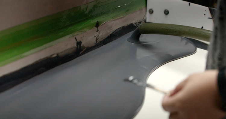
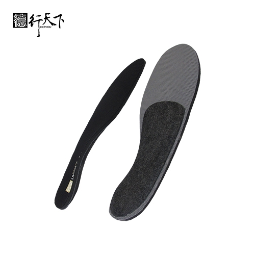
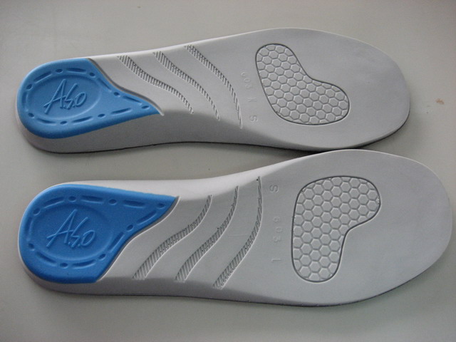
At GuangXin Industrial, our core strength lies in our deep expertise and versatility in insole and pillow manufacturing. We specialize in working with a wide range of materials, including PU (polyurethane), natural latex, and advanced graphene composites, to develop insoles and pillows that meet diverse performance, comfort, and health-support needs.
Whether it's cushioning, support, breathability, or antibacterial function, we tailor material selection to the exact requirements of each project-whether for foot wellness or ergonomic sleep products.
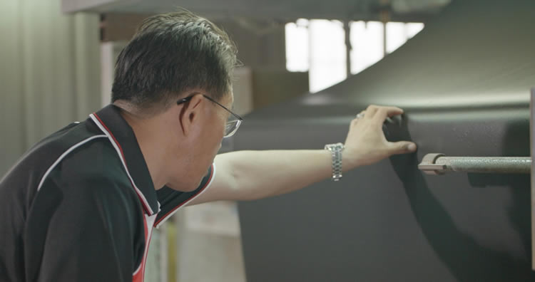
We provide end-to-end manufacturing capabilities under one roof—covering every stage from material sourcing and foaming, to precision molding, lamination, cutting, sewing, and strict quality control. This full-process control not only ensures product consistency and durability, but also allows for faster lead times and better customization flexibility.
With our flexible production capacity, we accommodate both small batch custom orders and high-volume mass production with equal efficiency. Whether you're a startup launching your first insole or pillow line, or a global brand scaling up to meet market demand, GuangXin is equipped to deliver reliable OEM/ODM solutions that grow with your business.
Customization & OEM/ODM Flexibility
GuangXin offers exceptional flexibility in customization and OEM/ODM services, empowering our partners to create insole products that truly align with their brand identity and target market. We develop insoles tailored to specific foot shapes, end-user needs, and regional market preferences, ensuring optimal fit and functionality.
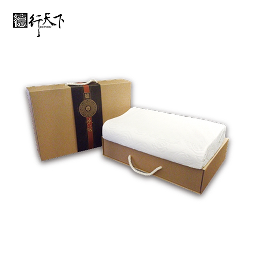
Our team supports comprehensive branding solutions, including logo printing, custom packaging, and product integration support for marketing campaigns. Whether you're launching a new product line or upgrading an existing one, we help your vision come to life with attention to detail and consistent brand presentation.
With fast prototyping services and efficient lead times, GuangXin helps reduce your time-to-market and respond quickly to evolving trends or seasonal demands. From concept to final production, we offer agile support that keeps you ahead of the competition.
Quality Assurance & Certifications
Quality is at the heart of everything we do. GuangXin implements a rigorous quality control system at every stage of production—ensuring that each insole meets the highest standards of consistency, comfort, and durability.
We provide a variety of in-house and third-party testing options, including antibacterial performance, odor control, durability testing, and eco-safety verification, to meet the specific needs of our clients and markets.
Our products are fully compliant with international safety and environmental standards, such as REACH, RoHS, and other applicable export regulations. This ensures seamless entry into global markets while supporting your ESG and product safety commitments.
ESG-Oriented Sustainable Production
At GuangXin Industrial, we are committed to integrating ESG (Environmental, Social, and Governance) values into every step of our manufacturing process. We actively pursue eco-conscious practices by utilizing eco-friendly materials and adopting low-carbon production methods to reduce environmental impact.
To support circular economy goals, we offer recycled and upcycled material options, including innovative applications such as recycled glass and repurposed LCD panel glass. These materials are processed using advanced techniques to retain performance while reducing waste—contributing to a more sustainable supply chain.
We also work closely with our partners to support their ESG compliance and sustainability reporting needs, providing documentation, traceability, and material data upon request. Whether you're aiming to meet corporate sustainability targets or align with global green regulations, GuangXin is your trusted manufacturing ally in building a better, greener future.
Let’s Build Your Next Insole Success Together
Looking for a reliable insole manufacturing partner that understands customization, quality, and flexibility? GuangXin Industrial Co., Ltd. specializes in high-performance insole production, offering tailored solutions for brands across the globe. Whether you're launching a new insole collection or expanding your existing product line, we provide OEM/ODM services built around your unique design and performance goals.
From small-batch custom orders to full-scale mass production, our flexible insole manufacturing capabilities adapt to your business needs. With expertise in PU, latex, and graphene insole materials, we turn ideas into functional, comfortable, and market-ready insoles that deliver value.
Contact us today to discuss your next insole project. Let GuangXin help you create custom insoles that stand out, perform better, and reflect your brand’s commitment to comfort, quality, and sustainability.
🔗 Learn more or get in touch:
🌐 Website: https://www.deryou-tw.com/
📧 Email: shela.a9119@msa.hinet.net
📘 Facebook: facebook.com/deryou.tw
📷 Instagram: instagram.com/deryou.tw
Custom graphene foam processing factory Taiwan
Are you looking for a trusted and experienced manufacturing partner that can bring your comfort-focused product ideas to life? GuangXin Industrial Co., Ltd. is your ideal OEM/ODM supplier, specializing in insole production, pillow manufacturing, and advanced graphene product design.
With decades of experience in insole OEM/ODM, we provide full-service manufacturing—from PU and latex to cutting-edge graphene-infused insoles—customized to meet your performance, support, and breathability requirements. Our production process is vertically integrated, covering everything from material sourcing and foaming to molding, cutting, and strict quality control.China graphene sports insole ODM
Beyond insoles, GuangXin also offers pillow OEM/ODM services with a focus on ergonomic comfort and functional innovation. Whether you need memory foam, latex, or smart material integration for neck and sleep support, we deliver tailor-made solutions that reflect your brand’s values.
We are especially proud to lead the way in ESG-driven insole development. Through the use of recycled materials—such as repurposed LCD glass—and low-carbon production processes, we help our partners meet sustainability goals without compromising product quality. Our ESG insole solutions are designed not only for comfort but also for compliance with global environmental standards.China anti-bacterial pillow ODM design
At GuangXin, we don’t just manufacture products—we create long-term value for your brand. Whether you're developing your first product line or scaling up globally, our flexible production capabilities and collaborative approach will help you go further, faster.Pillow OEM for wellness brands Taiwan
📩 Contact us today to learn how our insole OEM, pillow ODM, and graphene product design services can elevate your product offering—while aligning with the sustainability expectations of modern consumers.Taiwan sustainable material ODM production base
Dogs extract words from continuous speech using similar computations and brain regions as humans do, a new study combining EEG and fMRI by researchers from the Department of Ethology, Eötvös Loránd University (Hungary) finds. Credit: Grzegorz Eliasiewicz Dogs extract words from continuous speech using similar computations and brain regions as humans do, a new study combining EEG and fMRI by researchers from the Department of Ethology, Eötvös Loránd University (Hungary) finds. This is the first demonstration of the capacity to use complex statistics to learn about word boundaries in a non-human mammal. Human infants can spot new words in a speech stream much before they learn what those words mean. To tell where a word ends and another one begins, infants make complex calculations to keep track of syllable patterning: syllables that usually appear together are probably words, and those that do not probably aren’t. A new brain imaging study by Hungarian researchers discovered that dogs may also recognize such complex regularities in speech. “Keeping track of patterns is not unique to humans: many animals learn from such regularities in the surrounding world, this is called statistical learning. What makes speech special is that its efficient processing requires complex computations. Dog under EEG experiment. Credit: Grzegorz Eliasiewicz To learn new words from continuous speech, it is not enough to count how often certain syllables occur together. It is much more efficient to calculate how probably those syllables occur together. This is exactly how humans, even 8-month-old infants, solve the seemingly difficult task of word segmentation: they calculate complex statistics about the probability of one syllable following the other,” explains Marianna Boros, one of the lead authors of the study, and a postdoctoral researcher at the Neuroethology of Communication Lab, Department of Ethology, Eötvös Loránd University. “Until now we did not know if any other mammal can also use such complex computations to extract words from speech. We decided to test family dogs’ brain capacities for statistical learning from speech. Dogs are the earliest domesticated animal species and probably the one we speak most often to. Still, we know very little about the neural processes underlying their word learning capacities.” “To find out what kind of statistics dogs calculate when they listen to speech, first we measured their electric brain activity using EEG,” says Lilla Magyari, the other lead author, postdoctoral researcher in the same research group, who had laid the methodological foundations of performing non-invasive electrophysiology on awake, untrained, cooperating dogs. “Interestingly, we saw differences in dogs’ brain waves for frequent compared to rare words. But even more surprisingly, we also saw brain wave differences for syllables that always occurred together compared to syllables that only occasionally did, even if total frequencies were the same. So it turns out that dogs keep track not only of simple statistics (the number of times a word occurs) but also of complex statistics (the probability that a word’s syllables occur together). This has never been seen in other non-human mammals before. It is exactly the kind of complex statistics human infants use to extract words from continuous speech.” To explore how similar the responsible brain regions behind this complex computational capacity in dogs are to those in humans, researchers also tested dogs using functional MRI. This test was also performed on awake, cooperating, unrestrained animals. For fMRI, dogs were previously trained to lay motionless for the time of the measurements. Dog before fMRI with a trainer. Credit: Grzegorz Eliasiewicz “We know that in humans both general learning-related and language-related brain regions participate in this process. And we found the same duality in dogs,” explains Boros. “Both a generalist and a specialist brain region seemed to be involved in statistical learning from speech, but the activation patterns were different in the two. The generalist brain region, the so-called basal ganglia, responded stronger to a random speech stream (where no words could be spotted using syllable statistics) than to a structured speech stream (where words were easy to spot just by computing syllable statistics). The specialist brain region, the so-called auditory cortex, that in humans plays a key role in statistical learning from speech, showed a different pattern: here we saw brain activity increase over time for the structured but not for the random speech stream. We believe that this activity increase is the trace word learning leaves on the auditory cortex.” “We now begin to understand that some computational and neural processes that are known to be instrumental for human language acquisition may not be unique to humans after all,” says Attila Andics, principal investigator of the Neuroethology of Communication Lab. “But we still don’t know how these human-analog brain mechanisms for word learning emerged in dogs. Do they reflect skills that developed by living in a language-rich environment, or during the thousands of years of domestication, or do they represent an ancient mammalian capacity? We see that by studying speech processing in dogs, even better dog breeds with different communication abilities, and other species living close to humans, we can trace back the origins of human specializations for speech perception.” Reference: “Neural processes underlying statistical learning for speech segmentation in dogs” by Marianna Boros, Lilla Magyari, Dávid Török, Anett Bozsik, Andrea Deme and Attila Andics, 29 October 2021, Current Biology. DOI: 10.1016/j.cub.2021.10.017 This research was funded by the Hungarian Academy of Sciences and Eötvös Loránd Research Network (’Lendület’ Program), the European Research Council (ERC) and the Ministry for Innovation and Technology.
Reconstruction of Brevirostruavis macrohyoideus with its mouth open to show its long tongue that was used to catch insects or obtain nectar from cone-bearing plants. Credit: IVPP A new fossil skeleton of an extinct species of bird from northeastern China that lived alongside dinosaurs 120 million years ago unexpectedly preserves a bony tongue that is nearly as long as its head. The skull is very well preserved, showing that it had a relatively short snout and small teeth, with extremely long and curved bones for the tongue (called the hyoid apparatus). Scientists from the Institute of Vertebrate Paleontology and Paleoanthropology (IVPP) of the Chinese Academy of Sciences and the University of Texas at Austin have named this bird Brevirostruavis macrohyoideus, which means “bird with a short snout and big tongue.” Their discovery was published in Journal of Anatomy on December 1, 2021. We learn quickly as children to stick out our tongues, but most reptiles and birds do not have large muscular tongues like humans. Birds instead have a set of rod-shaped elements made of bone and cartilage comprising the hyoid apparatus that sits in the floor of their mouth. In birds with larger tongues like ducks and parrots, they use their tongue to move food around in their mouth, get food into their mouth, and help to swallow food. Some birds today like hummingbirds and woodpeckers have a bony tongue as long or longer than their skulls. Photograph and drawing of the skull of the extinct Cretaceous enantiornithine bird Brevirostruavis macrohyoideus, with the curved bones of the long tongue highlighted in orange. Credit: IVPP This extinct short-snouted, big-tongued bird is the earliest example of a bird being able to stick its tongue out. Of course, this feature makes one wonder why this bird would be sticking its tongue out. The scientists hypothesized that the bird might have used this feature for catching insects in the same way that living woodpeckers use their tongues to get insects out of holes in bark, wood, and tree branches. Alternatively, the bird might have been feeding on pollen or nectar-like liquids from plants in the forest where it lived. No stomach contents were found with this skeleton. This short-snouted, big-tongued bird is part of an extinct group of birds called enantiornithines or “opposite” birds. They were the most successful group of birds during the Cretaceous Period (between 66 and 145 million years ago), with fossils found around the world. “We see a lot of variation in the size and shape of the skulls of enantiornithine birds and that probably reflects the great diversity of the foods they ate and how they caught their food. Now with this fossil, we see that it’s not just their skulls, but their tongues that also vary,” said Dr. WANG Min, co-author of the study. The researchers previously showed that these early birds had fairly rigid skulls like their dinosaur relatives. This feature set some evolutionary and functional restrictions on early birds. “Perhaps the only way for them to fundamentally change through evolution how they caught their food and what food they ate was to shorten their skull in this case and to make the tongue bones much longer,” said lead author Dr. LI Zhiheng. The long, curved hyoid apparatus in the fossil bird is made of bones called ceratobranchials. Living birds also have such bones in their hyoid, but it is the epibranchial bones, absent in early birds, that are very long in birds like woodpeckers. “Animals experiment evolutionarily with what they have available. This bird evolved a long tongue using the bones it inherited from its dinosaur ancestors, and living birds evolved longer tongues with the bones that they have. This situation demonstrates the power of evolution, with birds using two different evolutionary pathways to solve the same problem of making a long tongue to stick out of their mouths,” said co-author Dr. Thomas Stidham. Reference: “Novel evolution of a hyper-elongated tongue in a Cretaceous enantiornithine from China and the evolution of the hyolingual apparatus and feeding in birds” by Zhiheng Li, Min Wang, Thomas A. Stidham, Zhonghe Zhou and Julia Clarke, 1 December 2021, Journal of Anatomy. DOI: 10.1111/joa.13588
The ER’s network of tubules rearranges itself, appearing green at first and then magenta after 60 seconds. White indicates areas where the network remains in position. Credit: Laura Westrate Scientists apply principles of math and physics to unravel the mystery of how the endoplasmic reticulum, an organelle vital to cellular life, constantly reshapes and reorganizes itself. As a second-year Ph.D. student and physicist, Zuben Scott hadn’t thought much about the endoplasmic reticulum since learning about cell structures as a high school freshman. Then a potential graduate adviser, Elena Koslover, suggested he study it. She showed him images and videos captured under a microscope that revealed an intricate mesh. “It was very beautiful,” he says of the organelle better known as simply the ER. “It was shocking to me to see how this complex network could form within cells.” Scott was equally intrigued by the question posed by Koslover, an associate professor of physics at the University of California, San Diego. Although best known as the site where proteins are assembled and prepared for their functions, the ER does much more. For example, it produces certain hormones and components of the cell membrane, and stores calcium ions, which cells use to coordinate responses to stimuli. These molecules move through the ER’s elaborate structure, and Koslover, who studies transport in cells from a physics perspective, wanted to investigate how. To do so, they need to account for the ever-changing nature of this organelle. “Constantly, every minute, the ER is restructuring and shifting around,” Koslover says. Working with the same images, which were taken by Koslover’s collaborator Laura Westrate, Scott eventually devised a model to describe this continuous reconfiguration. This research, published recently in Proceedings of the National Academy of Sciences, uncovers the unique dynamics governing the ER’s evolution and addresses the long-standing mystery of just how this organelle sustains life at the cellular level, with implications for understanding disease. A new kind of cellular network Before he took on the ER, Scott, who has since joined Adrian Jacobo’s research group at the Chan Zuckerberg Biohub San Francisco as a scientist, had dabbled in biophysics. The summer before his senior year as a physics major at Reed College, he first encountered this interdisciplinary field through a data-analysis internship working on a super-resolution imaging technique, with Xiaolin Nan at Oregon Health & Science University. The experience piqued his interest, but also revealed how much biology he had to learn to complement his knowledge of physics. “Xiaolin told me his seven-year-old knew more biology than me,” Scott says. “But eventually, I got there.” In Koslover’s lab, Scott became the designated “ER person.” For his project, he focused on the tube-filled section of the organelle adjacent to the cell’s membrane known as the peripheral ER. (Another portion, composed of sheets stacked like the levels of a parking garage, enfolds the nucleus.) He approached the tubular ER as a network, a term in physics that describes a set of connected points — think users on a social media platform, the intersections of a city’s roads, or in the case of the ER, the junctions where its tiny tubes meet, three at a time. Cells contain other networks too. Scientists describe the cell’s internal skeleton and its energy-converting mitochondria this way. But the dynamics that define other cellular networks don’t apply to the peripheral ER. “The junctions actually slide,” says Greg Huber, a UC San Francisco biophysicist and coauthor on the PNAS paper, describing how the three-tube connection points respond to the forces transmitted through the network of tubules. But this movement resembles that of a liquid, not a solid, so “the physical material that makes up a junction at one time will contain different molecules at a later time.” Huber, who previously led Biohub SF’s Physical Biology and Biophysical Theory Group, had been working on his own model for the peripheral ER when he joined Scott and Koslover’s project. To describe its behavior, he had taken to calling it a “liquid network,” but notes that, unlike an everyday liquid, the tubular ER generates its own shape. It is its own container, he says. Simple model, complicated structure To visualize this dynamic network of tubules, Westrate, of Calvin University, took advantage of a property of cells from a line known as COS-7: when grown in culture, these cells spread out like fried eggs. Thick in the middle but thin at the edges, this distinctive shape squishes the peripheral ER into nearly two dimensions, simplifying the task of studying it with imaging tools. The time-lapse images Westrate captured show new tubules spontaneously branching from and connecting to the existing network or other parts of the cell. Simultaneously, tension causes junctions to slide, shrinking tubules and closing the rings they form — leading to a network of simultaneously growing and shrinking shapes. Scott and Samuel Steen, from Westrate’s group, defined these dynamics mathematically by counting new tubules and determining the average area of the polygons the tubules enclosed. With these measurements, they derived the model’s two parameters: tubule growth rate and the mobility of the junctions. Using these parameters, it predicts the structural features of the ER with no further fiddling. “The best part about this model,” Scott says, “is its simplicity.” The same rules would likely apply to a 3D model of the ER; however, a 3D model would also need to account for the tubules’ widths, and this measurement would determine whether a growing tube intersects an existing one, or passes it by, he says. But even in 2D, this mathematical representation helps explain how the ER can, as previous research has shown, move about to explore most of a cell’s interior, delivering proteins, calcium, and other molecules as needed. In their paper, the team calls the peripheral ER an “active liquid network,” a term Huber coined, to capture not only the liquid-like sliding of the junctions but also the growth of new tubules from existing ones. “We have contributed — I hate to say it because the term’s so overused — a new paradigm,” Huber says, and he suspects that there are other examples of active liquid networks within the specialized internal structures of cells. By offering insight on how cells function normally, the team’s model is relevant to understanding how things go awry in disease, particularly Alzheimer’s disease, amyotrophic lateral sclerosis, and spastic paraplegia, which studies have linked to changes in the ER’s shape. Their research could also have more general implications for ER dysfunction in numerous other conditions, including heart disease and diabetes. Exploring life through physics When Scott joined Jacobo’s Quantitative Tissue Morphogenesis group in October, he traded the ER for the neuromast, a sensory organ found on the sides of the zebrafish that the lab studies to explore the dynamics involved in arranging cells to form organs during development. He is also planning a collaboration with colleagues at Stanford to examine the processes that maintain the midsection of the tube that forms the gut in fruit flies. The zebrafish neuromast and Drosophila gut have little in common with each other, let alone the ER, but Scott believes physical principles can contribute to understanding how these diverse structures form. After his time immersed in the ER and as a new member of an experimental biology lab, Scott now describes himself as “slightly less inexperienced” in biology, a field in which he still feels somewhat like an outsider. “I constantly oscillate between states of taking biology for granted and being in awe of the complexity of living systems,” he says. Reference: “The endoplasmic reticulum as an active liquid network” by Zubenelgenubi C. Scott, Samuel B. Steen, Greg Huber, Laura M. Westrate and Elena F. Koslover, 11 October 2024, Proceedings of the National Academy of Sciences. DOI: 10.1073/pnas.2409755121
DVDV1551RTWW78V
High-performance graphene insole OEM Thailand 》trusted by clients across wellness, footwear, and bedding industriesSoft-touch pillow OEM service in Vietnam 》seamless coordination from idea to finished productChina anti-odor insole OEM service 》minimizing lead time, maximizing product value
下一則: Arch support insole OEM from Taiwan 》expert-level
- ODM pillow for sleep brands China 》designed for qu
- ODM service for ergonomic pillows Taiwan 》dedicate
- China OEM/ODM hybrid insole services 》helping your
- Graphene sheet OEM supplier factory Taiwan 》experi
- Custom foam pillow OEM in Indonesia 》the preferred
- Arch support insole OEM from Taiwan 》expert-level
限會員,要發表迴響,請先登入


