Introduction – Company Background
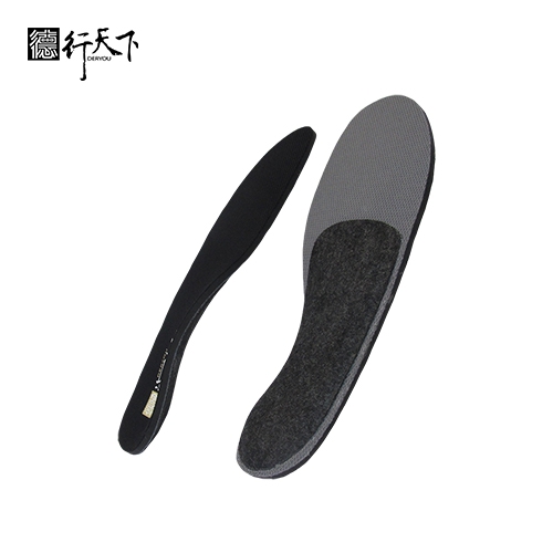
GuangXin Industrial Co., Ltd. is a specialized manufacturer dedicated to the development and production of high-quality insoles.
With a strong foundation in material science and footwear ergonomics, we serve as a trusted partner for global brands seeking reliable insole solutions that combine comfort, functionality, and design.
With years of experience in insole production and OEM/ODM services, GuangXin has successfully supported a wide range of clients across various industries—including sportswear, health & wellness, orthopedic care, and daily footwear.
From initial prototyping to mass production, we provide comprehensive support tailored to each client’s market and application needs.
At GuangXin, we are committed to quality, innovation, and sustainable development. Every insole we produce reflects our dedication to precision craftsmanship, forward-thinking design, and ESG-driven practices.
By integrating eco-friendly materials, clean production processes, and responsible sourcing, we help our partners meet both market demand and environmental goals.
Core Strengths in Insole Manufacturing
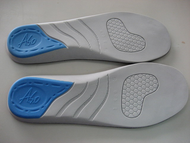
At GuangXin Industrial, our core strength lies in our deep expertise and versatility in insole and pillow manufacturing. We specialize in working with a wide range of materials, including PU (polyurethane), natural latex, and advanced graphene composites, to develop insoles and pillows that meet diverse performance, comfort, and health-support needs.
Whether it's cushioning, support, breathability, or antibacterial function, we tailor material selection to the exact requirements of each project-whether for foot wellness or ergonomic sleep products.
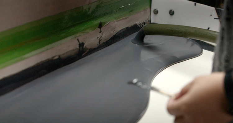
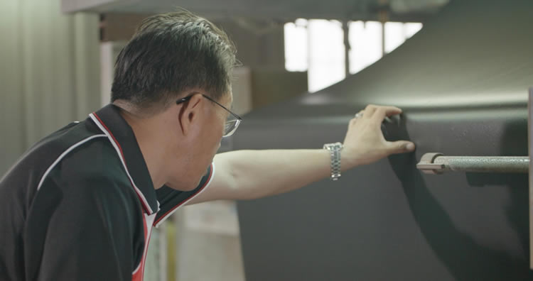
We provide end-to-end manufacturing capabilities under one roof—covering every stage from material sourcing and foaming, to precision molding, lamination, cutting, sewing, and strict quality control. This full-process control not only ensures product consistency and durability, but also allows for faster lead times and better customization flexibility.
With our flexible production capacity, we accommodate both small batch custom orders and high-volume mass production with equal efficiency. Whether you're a startup launching your first insole or pillow line, or a global brand scaling up to meet market demand, GuangXin is equipped to deliver reliable OEM/ODM solutions that grow with your business.
Customization & OEM/ODM Flexibility
GuangXin offers exceptional flexibility in customization and OEM/ODM services, empowering our partners to create insole products that truly align with their brand identity and target market. We develop insoles tailored to specific foot shapes, end-user needs, and regional market preferences, ensuring optimal fit and functionality.
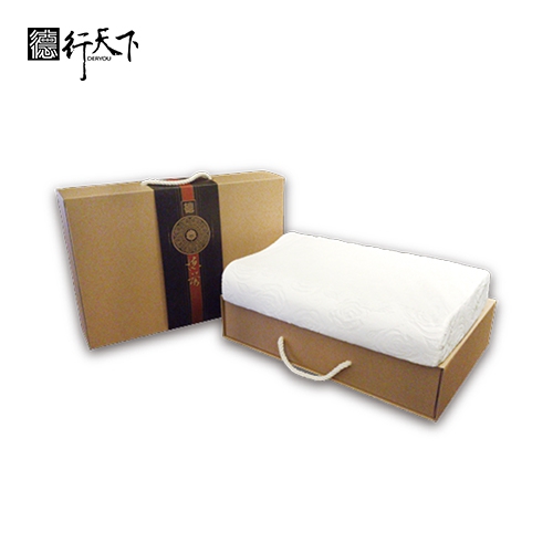
Our team supports comprehensive branding solutions, including logo printing, custom packaging, and product integration support for marketing campaigns. Whether you're launching a new product line or upgrading an existing one, we help your vision come to life with attention to detail and consistent brand presentation.
With fast prototyping services and efficient lead times, GuangXin helps reduce your time-to-market and respond quickly to evolving trends or seasonal demands. From concept to final production, we offer agile support that keeps you ahead of the competition.
Quality Assurance & Certifications
Quality is at the heart of everything we do. GuangXin implements a rigorous quality control system at every stage of production—ensuring that each insole meets the highest standards of consistency, comfort, and durability.
We provide a variety of in-house and third-party testing options, including antibacterial performance, odor control, durability testing, and eco-safety verification, to meet the specific needs of our clients and markets.
Our products are fully compliant with international safety and environmental standards, such as REACH, RoHS, and other applicable export regulations. This ensures seamless entry into global markets while supporting your ESG and product safety commitments.
ESG-Oriented Sustainable Production
At GuangXin Industrial, we are committed to integrating ESG (Environmental, Social, and Governance) values into every step of our manufacturing process. We actively pursue eco-conscious practices by utilizing eco-friendly materials and adopting low-carbon production methods to reduce environmental impact.
To support circular economy goals, we offer recycled and upcycled material options, including innovative applications such as recycled glass and repurposed LCD panel glass. These materials are processed using advanced techniques to retain performance while reducing waste—contributing to a more sustainable supply chain.
We also work closely with our partners to support their ESG compliance and sustainability reporting needs, providing documentation, traceability, and material data upon request. Whether you're aiming to meet corporate sustainability targets or align with global green regulations, GuangXin is your trusted manufacturing ally in building a better, greener future.
Let’s Build Your Next Insole Success Together
Looking for a reliable insole manufacturing partner that understands customization, quality, and flexibility? GuangXin Industrial Co., Ltd. specializes in high-performance insole production, offering tailored solutions for brands across the globe. Whether you're launching a new insole collection or expanding your existing product line, we provide OEM/ODM services built around your unique design and performance goals.
From small-batch custom orders to full-scale mass production, our flexible insole manufacturing capabilities adapt to your business needs. With expertise in PU, latex, and graphene insole materials, we turn ideas into functional, comfortable, and market-ready insoles that deliver value.
Contact us today to discuss your next insole project. Let GuangXin help you create custom insoles that stand out, perform better, and reflect your brand’s commitment to comfort, quality, and sustainability.
🔗 Learn more or get in touch:
🌐 Website: https://www.deryou-tw.com/
📧 Email: shela.a9119@msa.hinet.net
📘 Facebook: facebook.com/deryou.tw
📷 Instagram: instagram.com/deryou.tw
Vietnam custom neck pillow ODM
Are you looking for a trusted and experienced manufacturing partner that can bring your comfort-focused product ideas to life? GuangXin Industrial Co., Ltd. is your ideal OEM/ODM supplier, specializing in insole production, pillow manufacturing, and advanced graphene product design.
With decades of experience in insole OEM/ODM, we provide full-service manufacturing—from PU and latex to cutting-edge graphene-infused insoles—customized to meet your performance, support, and breathability requirements. Our production process is vertically integrated, covering everything from material sourcing and foaming to molding, cutting, and strict quality control.Indonesia high-end foam product OEM/ODM
Beyond insoles, GuangXin also offers pillow OEM/ODM services with a focus on ergonomic comfort and functional innovation. Whether you need memory foam, latex, or smart material integration for neck and sleep support, we deliver tailor-made solutions that reflect your brand’s values.
We are especially proud to lead the way in ESG-driven insole development. Through the use of recycled materials—such as repurposed LCD glass—and low-carbon production processes, we help our partners meet sustainability goals without compromising product quality. Our ESG insole solutions are designed not only for comfort but also for compliance with global environmental standards.ODM service for ergonomic pillows China
At GuangXin, we don’t just manufacture products—we create long-term value for your brand. Whether you're developing your first product line or scaling up globally, our flexible production capabilities and collaborative approach will help you go further, faster.China custom insole OEM supplier
📩 Contact us today to learn how our insole OEM, pillow ODM, and graphene product design services can elevate your product offering—while aligning with the sustainability expectations of modern consumers.Graphene sheet OEM supplier Taiwan
Scientists at UC San Francisco have created the first molecular-level, 3D image of how an odor molecule activates a human odorant receptor, paving the way for new insights into olfaction and its applications in fragrances and food science. The breakthrough enables researchers to potentially design new smells by understanding the interaction between scent molecules and odorant receptors. The First Molecular Images of Olfaction Have Opened the Door to Creating New Smells Scientists from UC San Francisco (UCSF) have accomplished a significant breakthrough in our understanding of olfaction by producing the first 3D image at the molecular level of how an odor molecule activates a human odorant receptor. This achievement is a crucial advancement toward unraveling the intricacies of the sense of smell. The findings, published in the journal Nature, are expected to rekindle interest in the science of smell, with far-reaching implications for fragrances, food science, and more. Odorant receptors, which are proteins situated on the surface of olfactory cells and bind to odor molecules, constitute half of the most diverse and extensive family of receptors in our bodies. A more comprehensive comprehension of them lays the groundwork for novel discoveries in a variety of biological processes. “This has been a huge goal in the field for some time,” said Aashish Manglik, MD, Ph.D., an associate professor of pharmaceutical chemistry and a senior author of the study. The dream, he said, is to map the interactions of thousands of scent molecules with hundreds of odorant receptors, so that a chemist could design a molecule and predict what it would smell like. “But we haven’t been able to make this map because, without a picture, we don’t know how odor molecules react with their corresponding odor receptors,” Manglik said. A Picture Paints the Scent of Cheese Smell involves about 400 unique receptors. Each of the hundreds of thousands of scents we can detect is made of a mixture of different odor molecules. Each type of molecule may be detected by an array of receptors, creating a puzzle for the brain to solve each time the nose catches a whiff of something new. “It’s like hitting keys on a piano to produce a chord,” said Hiroaki Matsunami, Ph.D., professor of molecular genetics and microbiology at Duke University and a close collaborator of Manglik. Matsunami’s work over the past two decades has focused on decoding the sense of smell. “Seeing how an odorant receptor binds an odorant explains how this works at a fundamental level.” To create that picture, Manglik’s lab used a type of imaging called cryo-electron microscopy (cryo-EM), that allows researchers to see atomic structure and study the molecular shapes of proteins. But before Manglik’s team could visualize the odorant receptor binding a scent molecule, they first needed to purify a sufficient quantity of the receptor protein. Odorant receptors are notoriously challenging, some say impossible, to make in the lab for such purposes. The Manglik and Matsunami teams looked for an odorant receptor that was abundant in both the body and the nose, thinking it might be easier to make artificially, and one that also could detect water-soluble odorants. They settled on a receptor called OR51E2, which is known to respond to propionate – a molecule that contributes to the pungent smell of Swiss cheese. But even OR51E2 proved hard to make in the lab. Typical cryo-EM experiments require a milligram of protein to produce atomic-level images, but co-first author Christian Billesbøelle, Ph.D., a senior scientist in the Manglik Lab, developed approaches to use only 1/100th of a milligram of OR51E2, putting the snapshot of receptor and odorant within reach. “We made this happen by overcoming several technical impasses that have stifled the field for a long time,” said Billesbøelle. “Doing that allowed us to catch the first glimpse of an odorant connecting with a human odorant receptor at the very moment a scent is detected.” This molecular snapshot showed that propionate sticks tightly to OR51E2 thanks to a very specific fit between odorant and receptor. The finding jibes with one of the duties of the olfactory system as a sentinel for danger. While propionate contributes to the rich, nutty aroma of Swiss cheese, on its own, its scent is much less appetizing. “This receptor is laser-focused on trying to sense propionate and may have evolved to help detect when food has gone bad,” said Manglik. Receptors for pleasing smells like menthol or caraway might instead interact more loosely with odorants, he speculated. Just a Whiff Along with employing a large number of receptors at a time, another interesting quality of the sense of smell is our ability to detect tiny amounts of odors that can come and go. To investigate how propionate activates this receptor, the collaboration enlisted quantitative biologist Nagarajan Vaidehi, Ph.D., at City of Hope, who used physics-based methods to simulate and make movies of how OR51E2 is turned on by propionate. “We performed computer simulations to understand how propionate causes a shape change in the receptor at an atomic level,” said Vaidehi. “These shape changes play a critical role in how the odorant receptor initiates the cell signaling process leading to our sense of smell.” The team is now developing more efficient techniques to study other odorant-receptor pairs and to understand the non-olfactory biology associated with the receptors, which have been implicated in prostate cancer and serotonin release in the gut. Manglik envisions a future where novel smells can be designed based on an understanding of how a chemical’s shape leads to a perceptual experience, not unlike how pharmaceutical chemists today design drugs based on the atomic shapes of disease-causing proteins. “We’ve dreamed of tackling this problem for years,” he said. “We now have our first toehold, the first glimpse of how the molecules of smell bind to our odorant receptors. For us, this is just the beginning.” Reference: “Structural basis of odorant recognition by a human odorant receptor” by Christian B. Billesbølle, Claire A. de March, Wijnand J. C. van der Velden, Ning Ma, Jeevan Tewari, Claudia Llinas del Torrent, Linus Li, Bryan Faust, Nagarajan Vaidehi, Hiroaki Matsunami and Aashish Manglik, 15 March 2023, Nature. DOI: 10.1038/s41586-023-05798-y Funding: This work was funded by the National Institutes of Health and the UCSF Program for Breakthrough Biomedical Research, funded in part by the Sandler Foundation. Cryo-EM equipment at UCSF is partially supported by NIH grants. For other funding, please see the paper.
Example of the morphological variation observed in the cranium of felids and nirmavids with species exhibiting both short and long upper canines. Credit: Narimane Chatar / University of Liège How did sabre-toothed tigers acquire their long upper canine teeth? In a groundbreaking study, an international team of scientists explored the evolutionary development of sabre teeth, revealing a complex continuum of cranial forms and accelerated evolutionary rates in sabre-toothed species. This study provides insights into the decline of these predators and broader evolutionary trends, enhancing our understanding of Earth’s past and the mechanisms of evolutionary convergence. Groundbreaking Study on Sabre Teeth Evolution Sabre teeth, those iconic elongated upper canine teeth, have long fascinated both scientists and the general public, notably because they have appeared several times in the fossil record, including two particularly well-known lineages of sabre-toothed tigers: the felids (the family of our domestic cats, lions, tigers, etc.) and the nimravids (a completely extinct family). However, the process by which these lineages acquired their elongated upper canines remains rather unclear. Skull and mandible of Eusmilus sicarius, a sabre-toothed nimravid scanned at Yale University Museum (Yale Paebody Museum, New Haven, USA.) Credit: N.Chatar/Université de Liège Detailed Analysis of Fossil and Modern Species Narimane Chatar, lead author of the study, who completed her doctorate at the EDDy Lab at the University of Liège and is now a post-doctoral fellow at UC Berkeley in the United States, has led an ambitious study to uncover the secrets of sabre tooth evolution. Using state-of-the-art 3D scanners and analytical methods, the team meticulously collected and analyzed data from a diverse set of current and extinct species. “We quantified the shape of 99 mandibles and 91 skulls, from different eras and continents, giving us a better understanding of the evolution of these animals,” explains Dr. Chatar. “Unlocking the secrets of sabre tooth evolution not only enriches our understanding of the Earth’s past, but also documents the mechanisms leading to evolutionary convergence,” says Professor Valentin Fischer, Director of the EDDyLab at ULiège. Surprising Findings in Sabre Tooth Evolution The study revealed some surprising results. The first is that rather than contrasting two distinct cranial morphologies in species with elongated upper canines and those with short teeth, there is instead a continuum of form linking the smallest present-day cats and their extinct sabre-toothed counterparts. “From a morphological point of view, the skull of a present-day small cat is just as strange and modified as that of a large sabre-toothed felid,” says Dr. Margot Michaud, a researcher at the University of French Guyana in Cayenne. These are therefore the two extremes of a continuum of forms that feline predators have seen evolve over geological time. “Our study suggests that what we often think of as examples of evolutionary patterns in textbooks are actually simplified for educational purposes. However, when we immerse ourselves in statistical analyses, we discover much more complex scenarios in these cases, as suggested by the results of our convergence tests,” explains Davide Tamagnini, post-doctoral researcher at the University of Rome La Sapienza. The second surprise concerns the path taken by evolution to produce sabre-toothed species. In fact, the team’s work has revealed that sabre-toothed species show faster rates of morphological evolution at the start of their evolutionary history than species with shorter canines. “Among other fascinating discoveries, we have shown that craniomandibular integration in sabre-toothed species is reduced, facilitating greater adaptability and diversification in the jaw and cranial morphology,” points out Margot Michaud. Thus, rapid morphological diversification and a fairly plastic skull have been identified as two key components that facilitated the emergence of elongated upper canines in both felids and nimravids. “As a result, there appears to be a common recipe for evolving into sabre-toothed feline-like predators,” says Dr. Chatar. Evolutionary Trends and Implications Finally, the team’s research highlighted the decline of sabre-toothed forms as well as the broader trends of feline-like predators over the course of their evolutionary history. Despite the relatively recent extinction of sabre-toothed forms ‘only’ a few thousand years ago, feline predators have in fact been in decline since the Miocene epoch (between -23 and -5 million years ago). “Some of these feline predators, particularly the sabre-toothed species, rapidly occupied fairly specialized niches, which made them more susceptible to extinction,” explains Dr. Tamagnini. This phenomenon, known as ‘ratchet’ or macroevolutionary ratchet, has been proposed as a potential driver for the decline of certain groups, where evolution favors the loss of early generalized forms, leading to the emergence of more specialized, but also more vulnerable, forms later in the history of the lineage. “Predators have their own evolutionary pathways and risks of extinction. Studying how ancient predators prospered and declined provides us with information about the possible futures of our ecosystems,” concludes Professor Fischer. Reference: “Evolutionary patterns of cat-like carnivorans unveil drivers of the sabertooth morphology” by Narimane Chatar, Margot Michaud, Davide Tamagnini and Valentin Fischer, 16 May 2024, Current Biology. DOI: 10.1016/j.cub.2024.04.055
Stanford University researchers discovered a new cellular pathway that clears misfolded proteins from the nucleus, which could be targeted for age-related disease therapies. The pathway involves communication between the nucleus and the cytoplasm, and the clearing process depends on a class of proteins that create small vesicles for transporting molecules. New pathway reveals how misfolded proteins are cleared from the nucleus, offering insights for neurodegenerative disease treatments. Misfolded proteins pose a threat to cellular health, as they interfere with normal functions and contribute to age-associated degenerative conditions such as Alzheimer’s, Parkinson’s, and Huntington’s diseases. The mechanisms by which cells eliminate these harmful proteins are not yet fully understood. A recent study, published on April 20 in Nature Cell Biology, reveals groundbreaking findings by Stanford University researchers. They uncovered a previously unidentified cellular pathway that facilitates the removal of misfolded proteins from the nucleus, where the cell’s DNA is stored, transcribed, and replicated. Maintaining the integrity of these processes is crucial for proper cellular function. This newly discovered pathway offers potential therapeutic targets for treating age-related diseases. To find the new pathway, researchers in the lab of Judith Frydman, the Donald Kennedy Chair in the School of Humanities and Sciences, integrated several genetic, imaging, and biochemical approaches to understand how yeast cells dealt with misfolded proteins. For the experiments, the team restricted misfolded proteins to either the nucleus or the cytoplasm – the area inside the cell but outside the nucleus. The team visually followed the fate of the misfolded proteins through live-cell imaging and super-resolution microscopy. A) A 3D reconstruction of a yeast cell engulfing cytoplasmic misfolded proteins (purple) inside of the degradation cellular machinery, or vacuole (gray). B) Super-resolution reconstructions showing nuclear misfolded proteins (green) being targeted to the degradation of cellular machinery through the nuclear-vacuolar junction (yellow). Credit: Fabián Morales-Polanco “The first exciting thing was that we actually found that there’s communication between the nucleus and the cytoplasm,” said Emily Sontag, the co-lead author of the paper and a former postdoctoral student in the Frydman Lab. “So they’re telling each other, ‘We both have a lot of misfolded proteins; let’s coordinate to send them here to this garbage dump so that they can be removed.’” The team identified the “garbage dump” site as the intersection of the nucleus and the vacuole – an organelle full of enzymes for degrading proteins – and showed that misfolded proteins in this “garbage dump” site are moved into the inside of the vacuole for degradation. They also showed that the pathway depends on a class of proteins used to create small vesicles for transporting molecules around cells. “Tying that particular family of proteins and this aspect of vesicle traffic biology to protein clearance gives us a new way to look at Alzheimer’s, Parkinson’s, Huntington’s – all these neurodegenerative diseases,” said Sontag. Shared ‘Garbage Dump’ Site for the Nucleus and the Cytoplasm Cells can deal with misfolded proteins in two ways: by refolding them or by eliminating them. A third option is to store them at a specific cellular location. “While the cell decides whether to refold or degrade proteins, it sequesters them into these membrane-less inclusions,” said Frydman, who is senior author of the paper. Inclusions are clusters of misfolded proteins that occur in both the cytoplasm and in the nucleus. The team found that the cellular machinery forms small misfolded-protein inclusions in different places within the nucleus and cytoplasm, like tiny garbage dumps, that then migrate toward the boundary between the nucleus and the vacuole, a bigger garbage dump. Eventually, the nuclear and cytoplasmic misfolded protein inclusions line up to face each other, with the nuclear envelope separating them. “The communication back and forth between the nucleus and the cytoplasm was not something we expected at all,” said Sontag. “Knowing that those two compartments can kind of work together to clear garbage from everywhere was really awesome.” “It shows that the management of misfolded proteins in the nucleus and the management of misfolded proteins in the cytoplasm are distinct but are coordinated,” said Frydman. “And what is really cool is that each compartment moves their misfolded proteins to the site where the nuclear envelope meets the vacuolar membrane.” From Dump Site to Degradation – a New Pathway The vacuole in yeast is equivalent to the lysosome in mammalian cells. It’s a membrane-bound organelle filled with enzymes that break down proteins – a recycling center for the cell. “This is not random,” said Fabián Morales-Polanco, the co-lead author of the paper and a postdoctoral scholar in the Frydman lab. “The cell is bringing inclusions to the same spot for a reason.” The team suspected that reason was to send the inclusions to the vacuole for degradation, but that raised further questions. It’s easy for cytoplasmic inclusions to enter the vacuole by autophagy – a process cells use to pull things from the cytoplasm into the vacuole or lysosome. But in the nucleus, inclusions are separated from the vacuole by the nuclear envelope. “Even though they come to the same spot, they don’t get into the vacuole by the same door,” said Morales-Polanco. To investigate the pathways of damaged proteins into the vacuole, the team blocked the proteasome – the other major protein clearance mechanism – and monitored the remaining protein clearance activity. They also created 3D images of the cells containing these misfolded protein inclusions using cryogenic soft X-ray tomography and fluorescence microscopy data. They found that the cytoplasmic inclusions did push into the vacuole, as expected. But the route for the nuclear inclusions was surprising. The nuclear inclusions budded straight from the nucleus into the vacuole at the junction of the two membranes. Using a series of genetic experiments, the team showed that ESCRT II/III and Vps4 proteins facilitated that budding-into-the-vacuole action. These proteins are known to cause membranes to bend and “bud,” or form new vesicles in other processes, but have not been studied as helping clear the nucleus of damaged proteins. They may be attractive therapy targets for misfolded protein diseases. Finally, using pH-sensitive tags, the team actually followed inclusions into the vacuole. “We were able to see these misfolded proteins entering into the vacuole and show this is really a new pathway,” said Morales-Polanco. An Eye on Aging The team did these experiments in yeast cells, which are easy to grow and quick to reproduce. One next step is to investigate whether this same pathway is used in mammalian cells to clear human disease-related proteins. Another next step is to define how the communication between the nucleus and cytosol happens along the pathway, and yet another is to see how the pathway is affected by aging. “There’s a lot of evidence that this process for dealing with misfolded proteins slows down with age,” said Sontag. “So, as time goes on, aged cells are not able to remove all that garbage as quickly or as efficiently, and misfolded proteins build up more and more inside the cell.” “We showed that nuclear and cytoplasmic quality control pathways communicate via the nuclear envelope, a structure that is impaired by aging and by neurodegenerative disease,” said Frydman. “Many progeria mutants, which cause premature aging, distort the nuclear envelope. This work really is a game changer in finally bringing a new way to understand, and hence cure, a wide range of terrible diseases that affect an increasingly aged population.” Reference: “Nuclear and cytoplasmic spatial protein quality control is coordinated by nuclear–vacuolar junctions and perinuclear ESCRT” by Emily M. Sontag, Fabián Morales-Polanco, Jian-Hua Chen, Gerry McDermott, Patrick T. Dolan, Daniel Gestaut, Mark A. Le Gros, Carolyn Larabell and Judith Frydman, 20 April 2023, Nature Cell Biology. DOI: 10.1038/s41556-023-01128-6 The study was funded by the National Institutes of Health, Way Klingler Faculty Development Awards from Marquette University, Pew Charitable Trusts, and the Gordon and Betty Moore Foundation.
DVDV1551RTWW78V
Graphene sheet OEM supplier Indonesia 》your competitive edge in product performance and speedTaiwan pillow OEM manufacturer 》empowering smart brands through better materials and processIndonesia insole ODM service provider 》helping your brand lead with innovation and integrity
限會員,要發表迴響,請先登入


