Introduction – Company Background
GuangXin Industrial Co., Ltd. is a specialized manufacturer dedicated to the development and production of high-quality insoles.
With a strong foundation in material science and footwear ergonomics, we serve as a trusted partner for global brands seeking reliable insole solutions that combine comfort, functionality, and design.
With years of experience in insole production and OEM/ODM services, GuangXin has successfully supported a wide range of clients across various industries—including sportswear, health & wellness, orthopedic care, and daily footwear.
From initial prototyping to mass production, we provide comprehensive support tailored to each client’s market and application needs.
At GuangXin, we are committed to quality, innovation, and sustainable development. Every insole we produce reflects our dedication to precision craftsmanship, forward-thinking design, and ESG-driven practices.
By integrating eco-friendly materials, clean production processes, and responsible sourcing, we help our partners meet both market demand and environmental goals.

Core Strengths in Insole Manufacturing
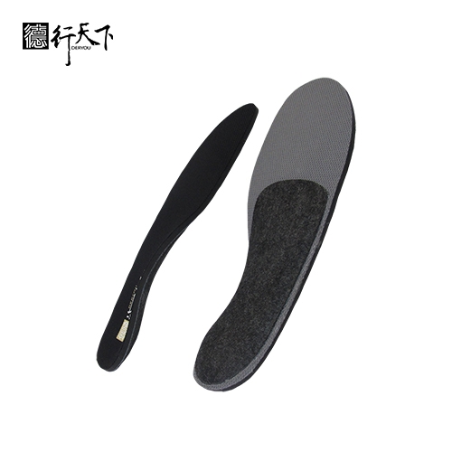
At GuangXin Industrial, our core strength lies in our deep expertise and versatility in insole and pillow manufacturing. We specialize in working with a wide range of materials, including PU (polyurethane), natural latex, and advanced graphene composites, to develop insoles and pillows that meet diverse performance, comfort, and health-support needs.
Whether it's cushioning, support, breathability, or antibacterial function, we tailor material selection to the exact requirements of each project-whether for foot wellness or ergonomic sleep products.
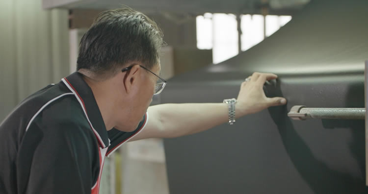
We provide end-to-end manufacturing capabilities under one roof—covering every stage from material sourcing and foaming, to precision molding, lamination, cutting, sewing, and strict quality control. This full-process control not only ensures product consistency and durability, but also allows for faster lead times and better customization flexibility.
With our flexible production capacity, we accommodate both small batch custom orders and high-volume mass production with equal efficiency. Whether you're a startup launching your first insole or pillow line, or a global brand scaling up to meet market demand, GuangXin is equipped to deliver reliable OEM/ODM solutions that grow with your business.
Customization & OEM/ODM Flexibility
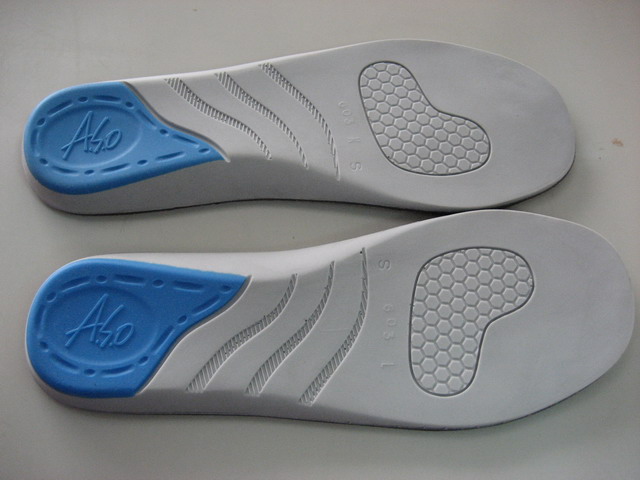
GuangXin offers exceptional flexibility in customization and OEM/ODM services, empowering our partners to create insole products that truly align with their brand identity and target market. We develop insoles tailored to specific foot shapes, end-user needs, and regional market preferences, ensuring optimal fit and functionality.
Our team supports comprehensive branding solutions, including logo printing, custom packaging, and product integration support for marketing campaigns. Whether you're launching a new product line or upgrading an existing one, we help your vision come to life with attention to detail and consistent brand presentation.
With fast prototyping services and efficient lead times, GuangXin helps reduce your time-to-market and respond quickly to evolving trends or seasonal demands. From concept to final production, we offer agile support that keeps you ahead of the competition.
Quality Assurance & Certifications
Quality is at the heart of everything we do. GuangXin implements a rigorous quality control system at every stage of production—ensuring that each insole meets the highest standards of consistency, comfort, and durability.
We provide a variety of in-house and third-party testing options, including antibacterial performance, odor control, durability testing, and eco-safety verification, to meet the specific needs of our clients and markets.
Our products are fully compliant with international safety and environmental standards, such as REACH, RoHS, and other applicable export regulations. This ensures seamless entry into global markets while supporting your ESG and product safety commitments.
ESG-Oriented Sustainable Production
At GuangXin Industrial, we are committed to integrating ESG (Environmental, Social, and Governance) values into every step of our manufacturing process. We actively pursue eco-conscious practices by utilizing eco-friendly materials and adopting low-carbon production methods to reduce environmental impact.
To support circular economy goals, we offer recycled and upcycled material options, including innovative applications such as recycled glass and repurposed LCD panel glass. These materials are processed using advanced techniques to retain performance while reducing waste—contributing to a more sustainable supply chain.
We also work closely with our partners to support their ESG compliance and sustainability reporting needs, providing documentation, traceability, and material data upon request. Whether you're aiming to meet corporate sustainability targets or align with global green regulations, GuangXin is your trusted manufacturing ally in building a better, greener future.
Let’s Build Your Next Insole Success Together
Looking for a reliable insole manufacturing partner that understands customization, quality, and flexibility? GuangXin Industrial Co., Ltd. specializes in high-performance insole production, offering tailored solutions for brands across the globe. Whether you're launching a new insole collection or expanding your existing product line, we provide OEM/ODM services built around your unique design and performance goals.
From small-batch custom orders to full-scale mass production, our flexible insole manufacturing capabilities adapt to your business needs. With expertise in PU, latex, and graphene insole materials, we turn ideas into functional, comfortable, and market-ready insoles that deliver value.
Contact us today to discuss your next insole project. Let GuangXin help you create custom insoles that stand out, perform better, and reflect your brand’s commitment to comfort, quality, and sustainability.
🔗 Learn more or get in touch:
🌐 Website: https://www.deryou-tw.com/
📧 Email: shela.a9119@msa.hinet.net
📘 Facebook: facebook.com/deryou.tw
📷 Instagram: instagram.com/deryou.tw
High-performance graphene insole OEM Vietnam
Are you looking for a trusted and experienced manufacturing partner that can bring your comfort-focused product ideas to life? GuangXin Industrial Co., Ltd. is your ideal OEM/ODM supplier, specializing in insole production, pillow manufacturing, and advanced graphene product design.
With decades of experience in insole OEM/ODM, we provide full-service manufacturing—from PU and latex to cutting-edge graphene-infused insoles—customized to meet your performance, support, and breathability requirements. Our production process is vertically integrated, covering everything from material sourcing and foaming to molding, cutting, and strict quality control.China ODM expert for comfort products
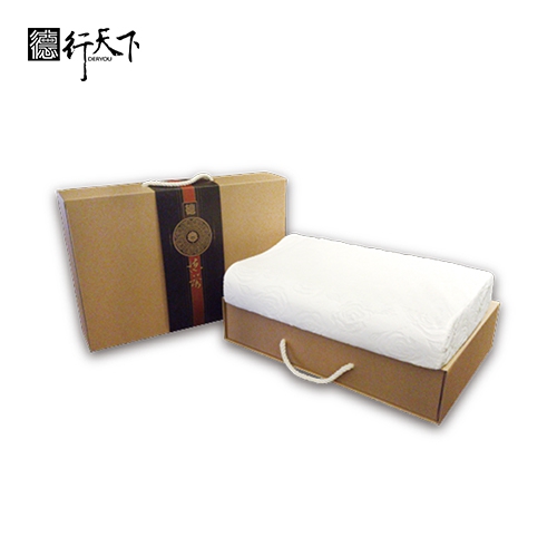
Beyond insoles, GuangXin also offers pillow OEM/ODM services with a focus on ergonomic comfort and functional innovation. Whether you need memory foam, latex, or smart material integration for neck and sleep support, we deliver tailor-made solutions that reflect your brand’s values.
We are especially proud to lead the way in ESG-driven insole development. Through the use of recycled materials—such as repurposed LCD glass—and low-carbon production processes, we help our partners meet sustainability goals without compromising product quality. Our ESG insole solutions are designed not only for comfort but also for compliance with global environmental standards.Private label insole and pillow OEM Thailand
At GuangXin, we don’t just manufacture products—we create long-term value for your brand. Whether you're developing your first product line or scaling up globally, our flexible production capabilities and collaborative approach will help you go further, faster.Vietnam OEM/ODM hybrid insole services
📩 Contact us today to learn how our insole OEM, pillow ODM, and graphene product design services can elevate your product offering—while aligning with the sustainability expectations of modern consumers.Soft-touch pillow OEM service in Indonesia
Researchers have developed a new genetic comparison technique that allows for a detailed study of the evolution of the human brain and face. New genetic comparison technique developed at Stanford enables meticulous study of evolution of the human brain and face. In separate studies, researchers compared gene regulation related to brain and face development in humans and chimpanzees using a new technique. In both cases, they discovered new genetic differences between these species. One of the best ways to study human evolution is by comparing us with nonhuman species that, evolutionarily speaking, are closely related to us. That closeness can help scientists narrow down precisely what makes us human, but that scope is so narrow it can also be extremely hard to define. To address this complication, researchers from Stanford University have developed a new technique for comparing genetic differences. Through two separate sets of experiments with this technique, the researchers discovered new genetic differences between humans and chimpanzees. They found a significant disparity in the expression of the gene SSTR2 – which modulates the activity of neurons in the cerebral cortex and has been linked, in humans, to certain neuropsychiatric diseases such as Alzheimer’s dementia and schizophrenia – and the gene EVC2, which is related to facial shape. The results were published March 17 in Nature and Nature Genetics, respectively. An image, from previous research, of human cortical spheroids derived in the lab of Sergiu Pașca, associate professor of psychiatry and behavioral sciences. Credit: Timothy Archibald “It’s important to study human evolution, not only to understand where we came from, but also why humans get so many diseases that aren’t seen in other species,” said Rachel Agoglia, a recent Stanford genetics graduate student who is lead author of the Nature paper. The Nature paper details the new technique, which involves fusing human and chimpanzee skin cells that had been modified to act like stem cells – highly malleable cells that can be prodded to transform into a variety of other cell types (albeit not a full organism). “These cells serve a very important specific purpose in this type of study by allowing us to precisely compare human and chimpanzee genes and their activities side-by-side,” said Hunter Fraser, associate professor of biology at Stanford’s School of Humanities and Sciences. Fraser is senior author of the Nature Genetics paper and co-senior author of the Nature paper with Sergiu Pașca, associate professor of psychiatry and behavioral sciences in the Stanford School of Medicine. Close Comparisons The Fraser lab is particularly interested in how the genetics of humans and other primates compare at the level of cis-regulatory elements, which affect the expression of nearby genes (located on the same DNA molecule, or chromosome). The alternative – called trans-regulatory factors – can regulate the expression of distant genes on other chromosomes elsewhere in the genome. Due to their broad effects, trans-regulatory factors (such as proteins) are less likely to differ among closely related species than cis-regulatory elements. But even when scientists have access to similar cells from humans and chimpanzees, there is a risk of confounding factors. For example, differences in the timing of development between species is a significant hurdle in studying brain development, explained Pașca. This is because human brains and chimpanzee brains develop at very different rates and there is no exact way to directly compare them. By housing human and chimpanzee DNA within the same cellular nucleus, scientists can exclude most confounding factors. For the initial experiments using these cells, Agoglia coaxed the cells into forming so-called cortical spheroids or organoids – a bundle of brain cells that closely mimics a developing mammalian cerebral cortex. The Pașca lab has been at the forefront of developing brain organoids and assembloids for the purpose of researching how the human brain is assembled and how this process goes awry in disease. “The human brain is essentially inaccessible at the molecular and cellular level for most of its development, so we introduced cortical spheroids to help us gain access to these important processes,” said Pașca, who is also the Bonnie Uytengsu and Family Director of Stanford Brain Organogenesis. As the 3D clusters of brain cells develop and mature in a dish, their genetic activity mimics what happens in early neurodevelopment in each species. Because the human and chimpanzee DNA are bound together in the same cellular environment, they are exposed to the same conditions and mature in parallel. Therefore, any observed differences in the genetic activity of the two can reasonably be attributed to actual genetic differences between our two species. Through studying brain organoids derived from the fused cells that were grown for 200 days, the researchers found thousands of genes that showed cis-regulatory differences between species. They decided to further investigate one of these genes – SSTR2 – which was more strongly expressed in human neurons and functions as a receptor for a neurotransmitter called somatostatin. In subsequent comparisons between human and chimpanzee cells, the researchers confirmed this elevated protein expression of SSTR2 in human cortical cells. Further, when the researchers exposed the chimpanzee cells and human cells to a small molecule drug that binds to SSTR2, they found that human neurons responded much more to the drug than the chimpanzee cells. This suggests a way by which the activity of human neurons in cortical circuits can be modified by neurotransmitters. Interestingly, this neuromodulatory activity may also be related to disease since SSTR2 has been shown to be involved in brain disease. “Evolution of the primate brain may have involved adding sophisticated neuromodulatory features to neural circuits, which under certain conditions can be perturbed and increase susceptibility to neuropsychiatric disease,” said Pașca. Fraser said these results are essentially “a proof of concept that the activity we’re seeing in these fused cells is actually relevant for cellular physiology.” Investigating Extreme Differences For the experiments published in Nature Genetics, the team coaxed their fused cells into cranial neural crest cells, which give rise to bones and cartilage in the skull and face, and determine facial appearance. “We were interested in these types of cells because facial differences are considered some of the most extreme anatomical differences between humans and chimps – and these differences actually affect other aspects of our behavior and evolution, like feeding, our senses, brain expansion, and speech,” said David Gokhman, a postdoctoral scholar in the Fraser lab and lead author of the Nature Genetics paper. “Also, the most common congenital diseases in humans are related to facial structure.” In the fused cells, the researchers identified a gene expression pathway that is much more active in the chimpanzee genes of the cells than in the human genes – with one specific gene, called EVC2, appearing to be six times more active in chimpanzees. Existing research has shown that people who have inactive EVC2 genes have flatter faces than others, suggesting that this gene could explain why humans have flatter faces than other primates. What’s more, the researchers determined that 25 observable facial features associated with inactive EVC2 are noticeably different between humans and chimpanzees – and 23 of those are different in the direction the researchers would have predicted, given lower EVC2 activity in humans. In follow-up experiments, where the researchers reduced the activity of EVC2 in mice, the rodents, too, developed flatter faces. Another Tool in the Toolbox This new experimental platform is not intended to replace existing cell comparison studies, but the researchers hope it will support many new findings about human evolution, and evolution in general. “Human development and the human genome have been very well studied,” said Fraser. “My lab is very interested in human evolution, but, because we can build on such a wealth of knowledge, this work can also reveal new insights into the process of evolution more broadly.” Looking forward, the Fraser lab is working on differentiating the fused cells into other cell types, such as muscle cells, other types of neurons, skin cells, and cartilage to expand their studies of uniquely human traits. The Pașca lab, meanwhile, is interested in investigating genetic dissimilarities related to astrocytes – large, multi-functional cells in the central nervous system often overlooked by scientists in favor of the flashier neurons. “While people often think about how neurons have evolved, we should not underestimate how astrocytes have changed during evolution. The size difference alone, between human astrocytes and astrocytes in other primates, is massive,” said Pașca. “My mentor, the late Ben Barres, called astrocytes ‘the basis of humanity’ and we absolutely think he was onto something.” References: “Primate cell fusion disentangles gene regulatory divergence in neurodevelopment” by Rachel M. Agoglia, Danqiong Sun, Fikri Birey, Se-Jin Yoon, Yuki Miura, Karen Sabatini, Sergiu P. Pașca and Hunter B. Fraser, 17 March 2021, Nature. DOI: 10.1038/s41586-021-03343-3 “Human–chimpanzee fused cells reveal cis-regulatory divergence underlying skeletal evolution” by David Gokhman, Rachel M. Agoglia, Maia Kinnebrew, Wei Gordon, Danqiong Sun, Vivek K. Bajpai, Sahin Naqvi, Coral Chen, Anthony Chan, Chider Chen, Dmitri A. Petrov, Nadav Ahituv, Honghao Zhang, Yuji Mishina, Joanna Wysocka, Rajat Rohatgi and Hunter B. Fraser, 17 March 2021, Nature Genetics. DOI: 10.1038/s41588-021-00804-3 Additional Stanford co-authors for the Nature paper are former research assistant Danqiong Sun, postdoctoral scholar Fikri Birey, senior research scientist Se-Jin Yoon, postdoctoral scholar Yuki Miura, and former research associate Karen Sabatini. This work was funded by a Stanford Bio-X Interdisciplinary Initiatives Seed Grant, the National Institutes of Health, the Department of Defense, the Stanford Center for Computational, Evolutionary and Human Genomics, the Stanford Medicine’s Dean’s Fellowship, MCHRI, the American Epilepsy Society, the Stanford Wu Tsai Neurosciences Institute’s Big Idea Grants on Brain Rejuvenation and Human Brain Organogenesis, the Kwan Research Fund, the New York Stem Cell Robertson Investigator Award, and the Chan Zuckerberg Ben Barres Investigator Award. Additional Stanford co-authors for the Nature Genetics paper are graduate student Maia Kinnebrew; former undergraduate Wei Gordon; former technician Danqiong Sun; postdoctoral research fellows Vivek Bajpai and Sahin Naqvi; Dmitri Petrov, the Michelle and Kevin Douglas Professor in the School of Humanities and Sciences; Joanna Wysocka, the Lorry Lokey Professor and professor of developmental biology; and Rajat Rohatgi, associate professor of biochemistry and of medicine. Researchers from University of California, San Francisco; University of Michigan, Ann Arbor; Yerkes National Primate Research Center; Emory University School of Medicine; and University of Pennsylvania are also co-authors. This work was funded by the Human Frontier, Rothschild and Zuckerman fellowships, and the National Institutes of Health. Fraser is a member of Stanford Bio-X, the Maternal & Child Health Research Institute (MCHRI), and the Stanford Cancer Institute. Pașca is a member of Stanford Bio-X, MCHRI and the Wu Tsai Neurosciences Institute, and a faculty fellow of Stanford ChEM-H.
Researchers are developing a detailed wiring diagram of the motor circuits in fruit flies’ central nervous systems. This connectome reveals the complex coordination between the nerves controlling leg and wing movements. Insights from recent studies highlight the complexity of motor neurons and their role in diverse movements like flying and walking, providing a basis for further research on neural circuit functionality. (Artist’s concept.) Credit: SciTechDaily Studies on fruit flies are shedding light on the complex neural coordination of movements, enhancing our understanding of motor neuron functionality. Scientists are developing a wiring diagram of the motor circuits in fruit flies’ central nervous system that control their muscles. This diagram, which is called a connectome, has already provided insights into the complex coordination between the nerves controlling leg and wing movements. Complexity in Simple Creatures While fruit flies seem like simple creatures, the researchers said their motor system contains “an unexpected level of complexity.” “A typical fly motor neuron receives thousands of synapses from hundreds of presynaptic premotor neurons,” the scientists observed. “This number is on par with the scale of synaptic integration in pyramidal cells of the rodent cortex.” An anatomical reconstruction of motor neurons that control muscles of the fruit fly leg and wing. Credit: Tyler Sloan/Quorometrix Studio New Studies on Motor Coordination Two new papers published in the scientific journal Nature have revealed the latest findings in this area, advancing our understanding of how the central nervous system in animals coordinates individual muscles to facilitate a variety of behaviors. Animation of the anatomical reconstruction of various nervous system structures involved in take-off and flight in a female fruit fly. Motor Neuron Efficiency and Adaptability Fruit flies use their legs for numerous activities such as leaping, walking, grooming, fighting, and courtship. They can also adapt their gait to navigate terrains like house plants, walls, damp surfaces, ceilings – and even insect-scale treadmills. All such movements, from postural reflexes that enable a fly to hold its position steady, to traversing obstacles or changing flight direction, originate through electrical signals from motor neurons. These signals are conducted through threadlike projections from the motor neuron to stimulate muscles. A fly’s six legs are managed by just 60 to 70 motor neurons, the researchers pointed out. In a cat, they noted, about 600 motor neurons supply a single feline calf muscle. Only 29 motor neurons govern the power and steering muscles of a fruit fly wing. In comparison, a hummingbird’s pectoral muscle is supplied by 2,000 motor neurons. Although the fly’s motor neurons are few, it performs remarkable aerial and terrestrial feats. An anatomical reconstruction of the ventral nerve cord of a female fruit fly. Credit: Tyler Sloan/Quorometrix Studio Wiring Logic of Premotor Circuits The scientists explained that motor units are composed of a single motor neuron and the muscle fibers that it can excite. Various motor units, activated in different combinations and sequences, collaborate to achieve a myriad of movement behaviors. The scientists in the two studies were interested in the wiring logic of premotor circuits. They wanted to understand how a fly’s nervous system coordinates motor units to accomplish varied tasks. Detailed Mapping and Synaptic Architecture One of the studies employed automated tools, machine learning, cell-type annotation, and electron microscopy to identify 14,600 neuronal cell bodies and about 45 million synapses (signal-transmission junctions) in the ventral nerve cord of a female fruit fly. The ventral nerve cord in flies is analogous to the spinal cord in vertebrates. The scientists subsequently applied deep learning to automatically reconstruct the anatomy of the neurons and their connections throughout the female fly. Motor Neurons and Muscle Activation in Flight The researchers used sophisticated methods to map the muscles targeted by leg and wing motor neurons. They determined which motor neurons in the female adult nerve cord connectome connect to individual muscles in the front leg and wing. From there, they created an atlas of the circuits that coordinate the fly’s leg and wing movements during take-off and flight motor initiation. To get into the air, the fly’s middle legs extend to jump and its front legs flex for departure. This is very roughly like a taxiing airliner retracting its wheels after leaving the ground or a wading heron tucking its spindly legs to keep them out of the way as it rushes into the sky. The scientists also found that some muscle fibers in adult flies are innervated by several motor neurons. This also occurs in the larval stage of the fruit fly and locusts. While some mammals have multiple innervations of nerve fibers as newborns, these usually disappear by adulthood. Multiple innervations might offer more flexibility and explain why an insect’s limbs can operate with precision despite having so few motor neurons. Functional and Evolutionary Insights from Fly Connectomics The scientists also examined the fly’s wing motor system, which has roughly three sections grouped by function: powering the wing flapping, steering the insect, and adjusting wing motion. The investigation of the connectivity of the premotor neurons enabled the researchers to compare the organization of premotor circuits for two types of limbs. The leg and the wing in fruit flies each have a distinct evolution and biomechanics. Implications and Future Directions of Connectome Research Connectomes are allowing scientists to produce new theories on how neural circuits function, and to debunk some false notions. The scientists mentioned that the recent community effort to develop the fruit fly connectome has led to one of the first synapse-level wiring diagrams for any limbed animal. They hope that additional connectomes will allow researchers to compare neural wiring across individuals. The anticipated reconstruction of a male fruit fly central nerve cord might illuminate differences between sexes. References: “Connectomic reconstruction of a female Drosophila ventral nerve cord” by Anthony Azevedo, Ellen Lesser, Jasper S. Phelps, Brandon Mark, Leila Elabbady, Sumiya Kuroda, Anne Sustar, Anthony Moussa, Avinash Khandelwal, Chris J. Dallmann, Sweta Agrawal, Su-Yee J. Lee, Brandon Pratt, Andrew Cook, Kyobi Skutt-Kakaria, Stephan Gerhard, Ran Lu, Nico Kemnitz, Kisuk Lee, Akhilesh Halageri, Manuel Castro, Dodam Ih, Jay Gager, Marwan Tammam, Sven Dorkenwald, Forrest Collman, Casey Schneider-Mizell, Derrick Brittain, Chris S. Jordan, Michael Dickinson, Alexandra Pacureanu, H. Sebastian Seung, Thomas Macrina, Wei-Chung Allen Lee and John C. Tuthill, 26 June 2024, Nature. DOI: 10.1038/s41586-024-07389-x “Synaptic architecture of leg and wing premotor control networks in Drosophila” by Ellen Lesser, Anthony W. Azevedo, Jasper S. Phelps, Leila Elabbady, Andrew Cook, Durafshan Sakeena Syed, Brandon Mark, Sumiya Kuroda, Anne Sustar, Anthony Moussa, Chris J. Dallmann, Sweta Agrawal, Su-Yee J. Lee, Brandon Pratt, Kyobi Skutt-Kakaria, Stephan Gerhard, Ran Lu, Nico Kemnitz, Kisuk Lee, Akhilesh Halageri, Manuel Castro, Dodam Ih, Jay Gager, Marwan Tammam, Sven Dorkenwald, Forrest Collman, Casey Schneider-Mizell, Derrick Brittain, Chris S. Jordan, Thomas Macrina, Michael Dickinson, Wei-Chung Allen Lee and John C. Tuthill, 26 June 2024, Nature. DOI: 10.1038/s41586-024-07600-z The research was supported by a Searle Scholar Award, Klingenstein-Simons Fellowship, Pew Biomedical Scholar Award, McKnight Scholar Award, Sloan Research Fellowship, New York Stem Cell Foundation, University of Washington Innovation Award, Genise Goldenson Award, National Institutes of Health Grants U19NS104655, RO1MH177808.
Researchers from the UK’s Medical Research Council Research Institutes have unraveled a longstanding mystery in DNA repair mechanisms, potentially enhancing cancer treatments. Their study revealed how the FANCD2-FANCI protein complex detects and initiates the repair of DNA cross-links, utilizing advanced imaging techniques to visualize this process at the molecular level. Researchers from the LMS and LMB have discovered how the D2-I protein complex identifies and repairs DNA damage, a breakthrough that promises to enhance cancer treatments by improving our understanding of DNA repair pathways. This collaboration could pave the way for more effective therapies by targeting the mechanisms that cancer cells use to resist treatment. A collaboration between researchers at the UK’s two core-funded Medical Research Council Institutes—the Laboratory of Medical Sciences (LMS) in London and the Laboratory of Molecular Biology (LMB) in Cambridge—has unraveled a decades-old mystery, potentially leading to improved cancer treatments in the future. The work, which uncovered the basic mechanism of how one of our most vital DNA repair systems recognizes DNA damages and initiates their repair, has eluded researchers for many years. Using cutting-edge imaging techniques to visualize how these DNA repair proteins move on a single molecule of DNA, and electron microscopy to capture how they “lock-on” to specific DNA structures, this research opens the way to more effective cancer treatments. The collaboration between the laboratories of Professor David Rueda (LMS) and Dr Lori Passmore (LMB) has been a brilliant example of how #teamscience can bear fruitful results and underscores the importance of these two institutes in driving forward research that unlocks the fundamental mechanisms of biology which will underpin the future translation of that work into improvements in human health. A single molecule of DNA (not directly visible) is captured using microscopic beads (the large circles). Each of the red, green, or yellow dots moving between the beads represents a FANCD2I-FANCI protein complex sliding along the DNA molecule, monitoring it for damage. Credit: MRC Laboratory of Medical Sciences Unraveling the DNA Repair Mechanism The researchers were working on a DNA repair pathway, known as the Fanconi Anaemia [FA] pathway, which was identified more than twenty years ago. DNA is constantly damaged throughout our lives by environmental factors including UV light from the sun, alcohol use, smoking, pollution, and exposure to chemicals. One way in which DNA becomes damaged is when it is “cross-linked”, which stops it being able to replicate and express genes normally. In order to replicate itself and to read and express genes, the two strands of the DNA double helix first has to unzip into single strands. When DNA is cross-linked, the “nucleotides” (the “steps” in the double-helix ladder of DNA) of the two strands become stuck together, preventing this unzipping. The accumulation of DNA damages including cross-linking can lead to cancer. The FA pathway is active throughout our lives and identifies these damages and repairs them on an ongoing basis. Individuals who have mutations that make this pathway less effective are far more susceptible to cancers. Although the proteins involved in the FA pathway were discovered some time ago, a mystery remained over how they identified the cross-linked DNA and started the process of DNA repair. The team from the MRC LMS sister institution, the LMB in Cambridge, led by Lori Passmore, had previously identified that the FANCD2-FANCI (D2-I) protein complex, which acts in one of the first steps of the FA pathway, clamps onto DNA, thereby initiating DNA repair at crosslinks. However, key questions remained: how does D2-I recognize crosslinked DNA, and why is the D2-I complex also implicated in other types of DNA damage? The research, published in the journal Nature, used a combination of cutting-edge scientific techniques to show that the D2-I complex slides along the double-stranded DNA, monitoring its integrity, and has also elegantly visualized how it recognizes where to stop, allowing the proteins to move and lock together at that point to initiate DNA repair. Advanced Techniques Shed Light on Molecular Interactions Artur Kaczmarczyk and Korak Ray in David Rueda’s Single Molecule Imaging group, working with Pablo Alcón in Lori Passmore’s group, used a state-of-the-art microscopy technique known as “correlated optical tweezers and fluorescence imaging” to explore how the D2-I complex slides along a double-stranded DNA molecule. Using optical tweezers, they could catch a single DNA molecule between two beads, which allowed them to precisely manipulate the DNA and incubate it with chosen proteins. Using fluorescently labeled D2-I and single-molecule imaging, they observed how individual D2-I complexes bind to and slide along DNA, scanning the double helix. They discovered that rather than recognizing the crosslink between the two strands of DNA directly, the FA clamp instead stops sliding when it reaches a single-stranded DNA gap, a region where one of the two strands of DNA is missing. The video shows the FANCD2-FANCI complex clamping to DNA in order to repair it. Credit: MRC Laboratory of Medical Sciences, MRC Laboratory for Molecular Biology Using cryo-electron microscopy, a powerful technique which can visualize proteins at a molecular level, the researchers next determined the structures of the D2-I complex both in its sliding position and stalled at the junction between single-stranded and double-stranded DNA. This revealed that the contacts D2-I makes with this single-stranded–double-stranded DNA junction are distinct from the contacts it makes with double-stranded DNA alone. This allowed them to identify a specific portion of the FANCD2 protein, called the “KR helix” that they showed in their single-molecule imaging experiments is critical for recognizing and stalling at the single-stranded DNA gaps. Working with Guillaume Guilbaud and Julian Sale in the LMB’s PNAC Division, and Themos Liolios and Puck Knipscheer at the Hubrecht Institute, Netherlands, they further showed that the D2-I complex’s ability to stall at these junctions using the KR helix is critical for DNA repair by the FA pathway. When DNA normally replicates in our cells, it unzips the two DNA strands and copies each single strand. This creates a ‘replication fork’ where the original DNA strands are unwound and new double-stranded DNA is formed on each strand. However, when this fork reaches a DNA crosslink, the strands cannot be unzipped, stalling the usual DNA replication process. This stalled replication fork thus contains exposed single-stranded gaps where the DNA has been unwound but not replicated. This research has shown that it is these junctions between single- and double-stranded DNA at the stalled replication fork that the D2-I protein complex latches tightly onto. Implications for Cancer Treatment and Beyond Not only does this allow D2-I complex to bring other FA pathway proteins to the DNA crosslink to initiate repair, but it also anchors the remaining double-stranded DNA, protecting the stalled “replication fork” from enzymes in the cell that would chew up the exposed end of the DNA strand and further damage the DNA. This work has shown that it is DNA structures within the replication fork that stalls as a result of cross-linked DNA, rather than the cross-linked DNA itself, that triggers the D2-I complex to stop sliding and clamp on to DNA to initiate repair. These stalled replication forks appear in many types of DNA damage, explaining the broad role of the D2-I complex in other forms of DNA repair as well as via the FA pathway. Understanding the process of DNA repair, and, importantly, why it fails, holds huge importance as DNA damage is a key factor in many diseases. Critically, many cancer drugs, for example, Cisplatin, work by inducing such serious cellular damage to cancer cells that they stop dividing and die. In such cases, DNA repair pathways—such a vital physiological process in normal life—can be hijacked by cancer cells that use them to resist the effects of chemotherapy drugs. Understanding the mechanistic basis of the first step in the DNA repair pathway may lead to ways of sensitizing patients so that cancer drugs can be more effective in the future. Reference: “FANCD2–FANCI surveys DNA and recognizes double- to single-stranded junctions” by Pablo Alcón, Artur P. Kaczmarczyk, Korak Kumar Ray, Themistoklis Liolios, Guillaume Guilbaud, Tamara Sijacki, Yichao Shen, Stephen H. McLaughlin, Julian E. Sale, Puck Knipscheer, David S. Rueda and Lori A. Passmore, 31 July 2024, Nature. DOI: 10.1038/s41586-024-07770-w This work was funded by UKRI MRC, the Wellcome Trust, the European Research Council, and the EMBO.
DVDV1551RTWW78V
Memory foam pillow OEM factory Thailand 》built to serve both niche and mass production needsThailand insole ODM service provider 》craftsmanship meets efficiency for maximum valueChina high-end foam product OEM/ODM 》functional, flexible, and built for scale
下一則: China anti-odor insole OEM service 》where craftsma
限會員,要發表迴響,請先登入


