Introduction – Company Background
GuangXin Industrial Co., Ltd. is a specialized manufacturer dedicated to the development and production of high-quality insoles.
With a strong foundation in material science and footwear ergonomics, we serve as a trusted partner for global brands seeking reliable insole solutions that combine comfort, functionality, and design.
With years of experience in insole production and OEM/ODM services, GuangXin has successfully supported a wide range of clients across various industries—including sportswear, health & wellness, orthopedic care, and daily footwear.
From initial prototyping to mass production, we provide comprehensive support tailored to each client’s market and application needs.
At GuangXin, we are committed to quality, innovation, and sustainable development. Every insole we produce reflects our dedication to precision craftsmanship, forward-thinking design, and ESG-driven practices.
By integrating eco-friendly materials, clean production processes, and responsible sourcing, we help our partners meet both market demand and environmental goals.
Core Strengths in Insole Manufacturing
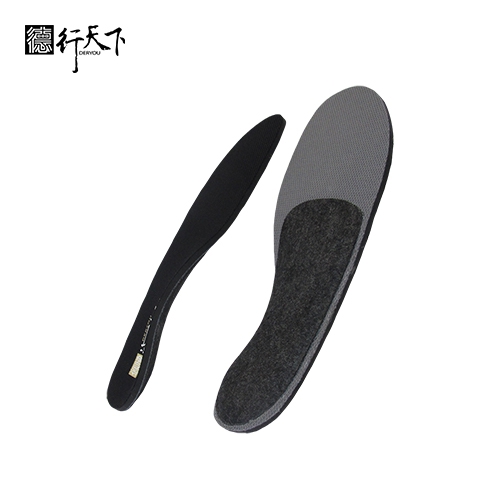
At GuangXin Industrial, our core strength lies in our deep expertise and versatility in insole and pillow manufacturing. We specialize in working with a wide range of materials, including PU (polyurethane), natural latex, and advanced graphene composites, to develop insoles and pillows that meet diverse performance, comfort, and health-support needs.
Whether it's cushioning, support, breathability, or antibacterial function, we tailor material selection to the exact requirements of each project-whether for foot wellness or ergonomic sleep products.
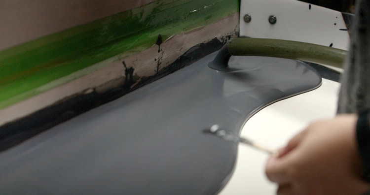
We provide end-to-end manufacturing capabilities under one roof—covering every stage from material sourcing and foaming, to precision molding, lamination, cutting, sewing, and strict quality control. This full-process control not only ensures product consistency and durability, but also allows for faster lead times and better customization flexibility.
With our flexible production capacity, we accommodate both small batch custom orders and high-volume mass production with equal efficiency. Whether you're a startup launching your first insole or pillow line, or a global brand scaling up to meet market demand, GuangXin is equipped to deliver reliable OEM/ODM solutions that grow with your business.
Customization & OEM/ODM Flexibility
GuangXin offers exceptional flexibility in customization and OEM/ODM services, empowering our partners to create insole products that truly align with their brand identity and target market. We develop insoles tailored to specific foot shapes, end-user needs, and regional market preferences, ensuring optimal fit and functionality.
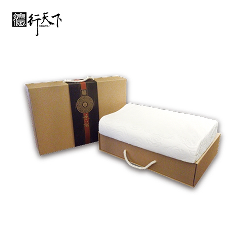
Our team supports comprehensive branding solutions, including logo printing, custom packaging, and product integration support for marketing campaigns. Whether you're launching a new product line or upgrading an existing one, we help your vision come to life with attention to detail and consistent brand presentation.
With fast prototyping services and efficient lead times, GuangXin helps reduce your time-to-market and respond quickly to evolving trends or seasonal demands. From concept to final production, we offer agile support that keeps you ahead of the competition.
Quality Assurance & Certifications
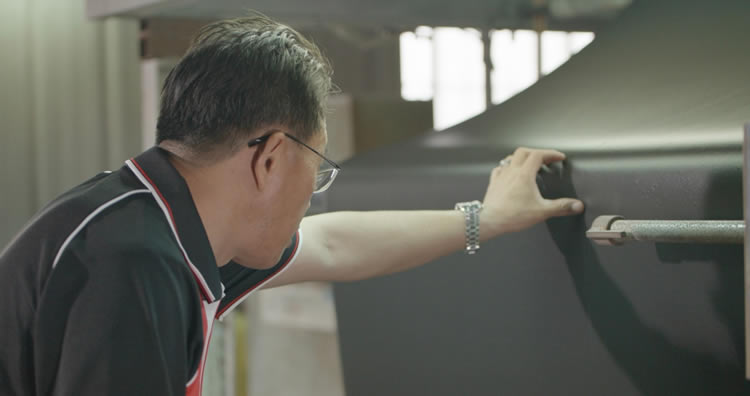
Quality is at the heart of everything we do. GuangXin implements a rigorous quality control system at every stage of production—ensuring that each insole meets the highest standards of consistency, comfort, and durability.
We provide a variety of in-house and third-party testing options, including antibacterial performance, odor control, durability testing, and eco-safety verification, to meet the specific needs of our clients and markets.
Our products are fully compliant with international safety and environmental standards, such as REACH, RoHS, and other applicable export regulations. This ensures seamless entry into global markets while supporting your ESG and product safety commitments.
ESG-Oriented Sustainable Production
At GuangXin Industrial, we are committed to integrating ESG (Environmental, Social, and Governance) values into every step of our manufacturing process. We actively pursue eco-conscious practices by utilizing eco-friendly materials and adopting low-carbon production methods to reduce environmental impact.
To support circular economy goals, we offer recycled and upcycled material options, including innovative applications such as recycled glass and repurposed LCD panel glass. These materials are processed using advanced techniques to retain performance while reducing waste—contributing to a more sustainable supply chain.
We also work closely with our partners to support their ESG compliance and sustainability reporting needs, providing documentation, traceability, and material data upon request. Whether you're aiming to meet corporate sustainability targets or align with global green regulations, GuangXin is your trusted manufacturing ally in building a better, greener future.
Let’s Build Your Next Insole Success Together
Looking for a reliable insole manufacturing partner that understands customization, quality, and flexibility? GuangXin Industrial Co., Ltd. specializes in high-performance insole production, offering tailored solutions for brands across the globe. Whether you're launching a new insole collection or expanding your existing product line, we provide OEM/ODM services built around your unique design and performance goals.
From small-batch custom orders to full-scale mass production, our flexible insole manufacturing capabilities adapt to your business needs. With expertise in PU, latex, and graphene insole materials, we turn ideas into functional, comfortable, and market-ready insoles that deliver value.
Contact us today to discuss your next insole project. Let GuangXin help you create custom insoles that stand out, perform better, and reflect your brand’s commitment to comfort, quality, and sustainability.
🔗 Learn more or get in touch:
🌐 Website: https://www.deryou-tw.com/
📧 Email: shela.a9119@msa.hinet.net
📘 Facebook: facebook.com/deryou.tw
📷 Instagram: instagram.com/deryou.tw
Eco-friendly pillow OEM manufacturer Indonesia
Are you looking for a trusted and experienced manufacturing partner that can bring your comfort-focused product ideas to life? GuangXin Industrial Co., Ltd. is your ideal OEM/ODM supplier, specializing in insole production, pillow manufacturing, and advanced graphene product design.
With decades of experience in insole OEM/ODM, we provide full-service manufacturing—from PU and latex to cutting-edge graphene-infused insoles—customized to meet your performance, support, and breathability requirements. Our production process is vertically integrated, covering everything from material sourcing and foaming to molding, cutting, and strict quality control.Graphene insole manufacturer in Indonesia
Beyond insoles, GuangXin also offers pillow OEM/ODM services with a focus on ergonomic comfort and functional innovation. Whether you need memory foam, latex, or smart material integration for neck and sleep support, we deliver tailor-made solutions that reflect your brand’s values.
We are especially proud to lead the way in ESG-driven insole development. Through the use of recycled materials—such as repurposed LCD glass—and low-carbon production processes, we help our partners meet sustainability goals without compromising product quality. Our ESG insole solutions are designed not only for comfort but also for compliance with global environmental standards.Vietnam neck support pillow OEM
At GuangXin, we don’t just manufacture products—we create long-term value for your brand. Whether you're developing your first product line or scaling up globally, our flexible production capabilities and collaborative approach will help you go further, faster.Vietnam insole OEM manufacturer
📩 Contact us today to learn how our insole OEM, pillow ODM, and graphene product design services can elevate your product offering—while aligning with the sustainability expectations of modern consumers.Ergonomic insole ODM support Thailand
This illustration of early Earth includes liquid water as well as magma seeping from the planet’s core due to a large impact. Scientists at NASA are investigating the chemistry that might have existed at this time in the planet’s history. Credit: Simone Marchi A specialized laboratory at JPL, designed to eliminate the chemical influence of modern organisms, allows scientists to investigate the chemistry that may have given rise to life. At NASA’s Jet Propulsion Laboratory’s Origins and Habitability Lab, you can find a world in a test tube. This is a simplified representation of the early Earth, reconstructed to resemble the conditions that existed on our planet around 4 billion years ago. Through this simulation, scientists are able to focus on the potential chemical reactions that occurred during that time, including those that could have played a crucial role in the development of life on Earth or could indicate the existence of life on other planets. Last year, researchers in JPL’s Origins and Habitability Lab simulated the chemistry of early Earth and carried out a key chemical reaction involved in metabolism, the process living organisms use to convert fuel (such as sunlight or food) into energy. Did Earth’s first life forms create energy with the same chemical reactions used by living organisms today? The first step to answering that question is finding out whether those reactions were even possible on early Earth. In living organisms, such reactions take place only inside a membrane (such as the protective wall of a living cell), which is just one reason why it’s an open question whether – and how – these reactions could have occurred before life formed. In JPL’s Origins and Habitability Lab, researchers use a sealed chamber to conduct experiments free of oxygen in an effort to replicate the chemistry of early Earth. Shown from left are lab co-lead Laurie Barge and researchers Jessica Weber and Laura Rodriguez. Credit: NASA/JPL-Caltech The lab’s work belongs to a discipline known as astrobiology: the study of the origins, evolution, distribution, and future of life in the universe. The threads all tie together, so trying to understand how life formed on Earth would also help scientists search for life elsewhere. In fact, in another study, the lab team looked into how understanding the origin of life on Earth can also help scientists interpret the appearance of organic molecules (the chemical basis for living things on Earth) that might be found on another planet or moon. But simulating the conditions found on Earth before life emerged is no easy task. Turning back the clock means taking into account how life has transformed our planet. Something in the Air There is essentially no place on Earth that’s not occupied by some form of life. Microorganisms can be found at the bottom of the ocean, in scalding hot geysers, and in rooms dedicated to removing those organisms. Life forms have also transformed our planet’s chemistry. One of the biggest challenges with trying to create pre-life conditions in the lab is dealing with the presence of oxygen. Largely absent from Earth’s atmosphere before life emerged, it is now ubiquitous because so many life forms produce it. As a result, all of the lab’s origin-of-life experiments have to be conducted inside an airtight box, with an airlock for putting items in or taking them out. In addition to test tubes containing chemicals, any instruments used to analyze those chemicals must fit within the box, so there are some experiments the team can’t do in this setting. Moreover, only one person can work in the box at a time, donning thick rubber gloves built into the sides of the container to move things around or use the equipment. Filters (which require regular cleaning) capture stray oxygen atoms. Even water has to go through a lengthy process to remove oxygen gas. “Science is all about repetition,” said JPL research scientist Laurie Barge, who co-leads the Origins and Habitability Lab. “We want to do experiments again and again, and that’s hard to do when you have to spend so much time making sure that not even a tiny bit of oxygen has crept into your test tube.” It took Barge and her team months to demonstrate that one chemical reaction involved in modern metabolism can take place under these early-Earth conditions. They plan to continue trying to simulate each step in the metabolism process and, at some point, they may find that a particular reaction can only occur inside a protective structure like a membrane. That might help narrow down when membranes became necessary in the emergence of life – a glimpse back in time. Testing Hypotheses on Other Worlds There’s another way that scientists could learn about the chemistry that took place, and potentially set the stage, for life on Earth: By studying a planet or moon with roughly the same raw ingredients that would have been found on early Earth. The location could be a lifeless moon in our own solar system or a planet around another star. Then Barge and her colleagues could test the ideas they’re investigating against an environment that isn’t constrained to the size of a glove box. “It would be very interesting to validate and check some of our laboratory results against results from another world,” said Jessica Weber, a JPL research scientist in the Origins and Habitability Lab who led the metabolism work. “Finding an environment like this would help us better re-create early Earth in our lab experiments, and that would get us closer to answering some of those big questions about life on our own planet and potentially on others.” References: “Determining the “Biosignature Threshold” for Life Detection on Biotic, Abiotic, or Prebiotic Worlds” by Laura M. Barge, Laura E. Rodriguez, Jessica M. Weber and Bethany P. Theiling, 13 April 2022, Astrobiology. DOI: 10.1089/ast.2021.0079 “Testing Abiotic Reduction of NAD+ Directly Mediated by Iron/Sulfur Minerals” by Jessica M. Weber, Bryana L. Henderson, Douglas E. LaRowe, Aaron D. Goldman, Scott M. Perl, Keith Billings and Laura M. Barge, 11 January 2022, Astrobiology. DOI: 10.1089/ast.2021.0035
By engineering “programmable” embryo-like models, scientists can now study early development without real embryos. Stem cells, guided by CRISPR, self-organize into structures that mirror natural processes, offering insights into fertility and genetic disorders. Credit: Ali Shariati/ UC Santa Cruz, edited Scientists have found a way to study early embryonic development without real embryos. Using CRISPR, they programmed stem cells to self-organize into structures mimicking early embryos. The cells show remarkable, collective behavior, almost as if they instinctively “know” what to do. This breakthrough allows researchers to explore genetic influences on development, reproductive challenges, and potential fertility treatments—all without using actual embryos. Unlocking the Earliest Mysteries of Life The earliest moments after fertilization, when a sperm cell meets an egg, remain one of biology’s greatest mysteries. Scientists from various fields are fascinated by how a single cell develops into a complex organism. In many animals, this transformation occurs inside the protective environment of the uterus, making direct observation difficult. As a result, researchers struggle to fully understand what can go wrong during early development and how external factors might prevent embryo formation. Engineering Embryos Without Embryos To overcome these challenges, scientists at UC Santa Cruz have engineered cellular models that replicate the first few days of embryonic development, without using actual embryos. Using CRISPR-based techniques, they guide stem cells to self-organize into “programmable” embryo-like structures, known as embryoids. These lab-grown cell assemblies are not true embryos but closely mimic key aspects of early development, providing a powerful tool for studying genetic and environmental influences on embryonic formation. Their findings were published today (March 20) in Cell Stem Cell, a leading journal in stem cell research. “We as scientists are interested in recreating and repurposing natural phenomena, such as formation of an embryo, in the dish to enable studies that are otherwise challenging to do with natural systems,” said Ali Shariati, assistant professor of biomolecular engineering and the study’s senior author. “We want to know how cells organize themselves into an embryo-like model, and what could go wrong when there are pathological conditions that prevent an animal from successfully developing.” Cell Co-Development: A More Natural Approach Shariati is an expert in stem cell engineering, a field that uses stem cells — unspecialized cells that can form any type of cell such as gut or brain cells — to study and solve biological and health problems. This project, led by UCSC postdoctoral scholar Gerrald Lodewijk and biomolecular engineering alumna and current Caltech graduate student Sayaka Kozuki, used mouse stem cells that are commonly grown in the lab to guide them to form basic building blocks of the embryo. Programmable cellular models of embryos, known as embryoids, allow scientists to mimic the first few day of embryonic development. Credit: Ali Shariati/ UC Santa Cruz CRISPR Technology: A Revolutionary Tool The team used a version of CRISPR technology known as an epigenome editor, which does not cut DNA but instead modifies how it is expressed. They targeted regions of the genome known to be involved in the development of an early embryo. This allowed them to control which genes were activated, and induce the creation of main types of cells needed for early development. “We use the stem cells, which are like a blank canvas, and use them to induce different cell types using our CRISPR tools,” Lodewijik said. This method had the advantage of allowing different cell types to “co-develop,” which more closely resembles the natural embryo formation than the chemical approaches other scientists have used to develop different cell types. “These cells co-develop together, just like they would in an actual embryo, and establish that history of being neighbors,” Shariati said. “We do not change their genome or expose them to specific signaling molecules, but rather activate the existing genes.” Self-Organizing Cells: A Remarkable Discovery The team found that 80% of the stem cells organize themselves into a structure that mimics the most basic form of an embryo after a few days, and most undergo gene activation that reflects the development process that occurs in living organisms. “The similarity is remarkable in the way the cells organize themselves, as well as the molecular composition,” Shariati said. “[The cells require] very little input from us — it’s as if the cells already know what to do, and we just give them a little bit of guidance.” The researchers observed that the cells showed a collective behavior in moving and organizing together. “Some of them start doing this rotational migration, almost like the collective behavior of birds or other species,” Shariati said. “Through this collective behavior and migration they can form these fascinating embryonic patterns.” “Programmable” Models for Developmental Research Having an accurate baseline model that reflects a living organisms’ early embryo could allow scientists to better study and learn how to treat developmental disorders or mutations. “These models have a more complete representation of what’s going on in early stages of development, and can capture the background,” Lodewijik said. The CRISPR programming not only allows the scientists to activate the genes at the beginning of the experimentation process, but also enables them to activate or modify genes important for other parts of development. This allows the embryo models to be “programmable,” meaning they can be relatively easily influenced with a high level of control to target and test the impact of multiple genes as the embryo model develops, illuminating which have deleterious effects when turned on or off. As an example, the researchers demonstrated how certain tissues form or are hindered during early development, but their methods could be used to study a wide range of genes and their cascading effects on the cell types. “I think this is the pioneering work of this study — the programmability and that we don’t rely on extrinsic factors to do this, but rather have a lot of control inside the cell,” Shariati said. The researchers are interested in how this approach might be used to study other species, allowing for a look into their embryo formation without ever using their actual embryos. A Window Into Fertility Challenges This research could allow for the study of the bottlenecks that lead reproduction to fail in early stages. Among mammals, humans have more reproduction challenges in that human embryos often fail to implant or establish the correct early organizational form. Understanding why this is the case could help make progress toward improving human fertility. Reference: “Self-organization of mouse embryonic stem cells into reproducible pre-gastrulation embryo models via CRISPRa programming” by Gerrald A. Lodewijk, Sayaka Kozuki, Clara J. Han, Benjamin R. Topacio, Seungho Lee, Lily Nixon, Abolfazl Zargari, Gavin Knight, Randolph Ashton, Lei S. Qi and S. Ali Shariati, 20 March 2025, Cell Stem Cell. DOI: 10.1016/j.stem.2025.02.015
Salk scientists developed a technique of using sound waves to control brain cells, dubbed sonogenetics, to selectively and noninvasively turn on groups of neurons. It was first used on worms and now has been used on mammalian cells. This technique could be a boon to science and medicine. Credit: Courtesy of the Salk Institute for Biological Studies Salk researchers pinpoint a sound-sensitive mammalian protein that lets them activate brain, heart or other cells with ultrasound. Salk scientists have engineered mammalian cells to be activated using ultrasound. The method, which the team used to activate human cells in a dish and brain cells inside living mice, paves the way toward non-invasive versions of deep brain stimulation, pacemakers and insulin pumps. The findings will be published in Nature Communications today (February 9, 2022). “Going wireless is the future for just about everything,” says senior author Sreekanth Chalasani, an associate professor in Salk’s Molecular Neurobiology Laboratory. “We already know that ultrasound is safe, and that it can go through bone, muscle, and other tissues, making it the ultimate tool for manipulating cells deep in the body.” Challenges and Discoveries in Protein Screening About a decade ago, Chalasani pioneered the idea of using ultrasonic waves to stimulate specific groups of genetically marked cells, and coined the term “sonogenetics” to describe it. In 2015, his group showed that, in the roundworm Caenorhabditis elegans, a protein called TRP-4 makes cells sensitive to low-frequency ultrasound. When the researchers added TRP-4 to C. elegans neurons that didn’t usually have it, they could activate these cells with a burst of ultrasound—the same sound waves used in medical sonograms. Neurons (magenta) in the mouse brain. The Chalasani lab made specific neurons express TRPA1 (white), so they can be activated by ultrasound. Credit: Salk Institute When the researchers tried adding TRP-4 to mammalian cells, however, the protein was not able to make the cells respond to ultrasound. A few mammalian proteins were reported to be ultrasound-sensitive, but none seemed ideal for clinical use. So Chalasani and his colleagues set out to search for a new mammalian protein that made cells highly ultrasound sensitive at 7 MHz, considered an optimal and safe frequency. “Our approach was different than previous screens because we set out to look for ultrasound-sensitive channels in a comprehensive way,” says Yusuf Tufail, a former project scientist at Salk and a co-first author of the new paper. TRPA1 Protein The researchers added hundreds of different proteins, one at a time, to a common human research cell line (HEK), which does not usually respond to ultrasound. Then, they put each cell culture under a setup that let them monitor changes to the cells upon ultrasound stimulation. Top from left: Sreekanth Chalasani and Corinne Lee-Kubli. Bottom from left: Marc Duque and Yusuf Tufail. Credit: Top: Salk Institute. Bottom from left: Marc Duque and Yusuf Tufail After screening proteins for more than a year, and working their way through nearly 300 candidates, the scientists finally found one that made the HEK cells sensitive to the 7 MHz ultrasound frequency. TRPA1, a channel protein, was known to let cells respond to the presence of noxious compounds and to activate a range of cells in the human body, including brain and heart cells. But Chalasani’s team discovered that the channel also opened in response to ultrasound in HEK cells. “We were really surprised,” says co-first author of the paper Marc Duque, a Salk exchange student. “TRPA1 has been well-studied in the literature but hasn’t been described as a classical mechanosensitive protein that you’d expect to respond to ultrasound.” To test whether the channel could activate other cell types in response to ultrasound, the team used a gene therapy approach to add the genes for human TRPA1 to a specific group of neurons in the brains of living mice. When they then administered ultrasound to the mice, only the neurons with the TRPA1 genes were activated. Expanding Sonogenetics Applications Beyond the Lab Clinicians treating conditions including Parkinson’s disease and epilepsy currently use deep brain stimulation, which involves surgically implanting electrodes in the brain, to activate certain subsets of neurons. Chalasani says that sonogenetics could one day replace this approach—the next step would be developing a gene therapy delivery method that can cross the blood-brain barrier, something that is already being studied. Perhaps sooner, he says, sonogenetics could be used to activate cells in the heart, as a kind of pacemaker that requires no implantation. “Gene delivery techniques already exist for getting a new gene—such as TRPA1—into the human heart,” Chalasani says. “If we can then use an external ultrasound device to activate those cells, that could really revolutionize pacemakers.” For now, his team is carrying out more basic work on exactly how TRPA1 senses ultrasound. “In order to make this finding more useful for future research and clinical applications, we hope to determine exactly what parts of TRPA1 contribute to its ultrasound sensitivity and tweak them to enhance this sensitivity,” says Corinne Lee-Kubli, a co-first author of the paper and former postdoctoral fellow at Salk. They also plan to carry out another screen for ultrasound sensitive proteins—this time looking for proteins that can inhibit, or shut off, a cell’s activity in response to ultrasound. Reference: “Sonogenetic control of mammalian cells using exogenous Transient Receptor Potential A1 channels” by Marc Duque, Corinne A. Lee-Kubli, Yusuf Tufail, Uri Magaram, Janki Patel, Ahana Chakraborty, Jose Mendoza Lopez, Eric Edsinger, Aditya Vasan, Rani Shiao, Connor Weiss, James Friend and Sreekanth H. Chalasani, 9 February 2022, Nature Communications. DOI: 10.1038/s41467-022-28205-y The other authors of the paper were Uri Magaram, Janki Patel, Ahana Chakraborty, Jose Mendoza Lopez, Eric Edsinger, Rani Shiao and Connor Weiss of Salk; and Aditya Vasan and James Friend of UC San Diego. The work was supported by the National Institutes of Health (R01MH111534, R01NS115591), Brain Research Foundation, Kavli Institute of Brain and Mind, Life Sciences Research Foundation, W.M. Keck Foundation (SERF), and the Waitt Advanced Biophotonics and GT3 Cores (which receive funding through NCI CCSG P30014195 and NINDSR24).
DVDV1551RTWW78V
Taiwan custom product OEM/ODM services 》committed to helping you create value through custom manufacturingThailand custom product OEM/ODM services 》where form meets function, every step of the wayLatex pillow OEM production in Thailand 》a manufacturing partner you can rely on for quality and delivery
下一則: Graphene cushion OEM factory in Vietnam 》driving y
限會員,要發表迴響,請先登入


