Introduction – Company Background
GuangXin Industrial Co., Ltd. is a specialized manufacturer dedicated to the development and production of high-quality insoles.
With a strong foundation in material science and footwear ergonomics, we serve as a trusted partner for global brands seeking reliable insole solutions that combine comfort, functionality, and design.
With years of experience in insole production and OEM/ODM services, GuangXin has successfully supported a wide range of clients across various industries—including sportswear, health & wellness, orthopedic care, and daily footwear.
From initial prototyping to mass production, we provide comprehensive support tailored to each client’s market and application needs.
At GuangXin, we are committed to quality, innovation, and sustainable development. Every insole we produce reflects our dedication to precision craftsmanship, forward-thinking design, and ESG-driven practices.
By integrating eco-friendly materials, clean production processes, and responsible sourcing, we help our partners meet both market demand and environmental goals.
Core Strengths in Insole Manufacturing
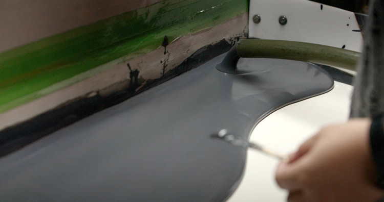
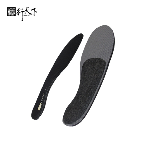
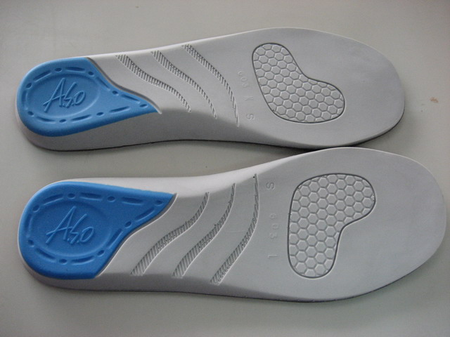
At GuangXin Industrial, our core strength lies in our deep expertise and versatility in insole and pillow manufacturing. We specialize in working with a wide range of materials, including PU (polyurethane), natural latex, and advanced graphene composites, to develop insoles and pillows that meet diverse performance, comfort, and health-support needs.
Whether it's cushioning, support, breathability, or antibacterial function, we tailor material selection to the exact requirements of each project-whether for foot wellness or ergonomic sleep products.
We provide end-to-end manufacturing capabilities under one roof—covering every stage from material sourcing and foaming, to precision molding, lamination, cutting, sewing, and strict quality control. This full-process control not only ensures product consistency and durability, but also allows for faster lead times and better customization flexibility.
With our flexible production capacity, we accommodate both small batch custom orders and high-volume mass production with equal efficiency. Whether you're a startup launching your first insole or pillow line, or a global brand scaling up to meet market demand, GuangXin is equipped to deliver reliable OEM/ODM solutions that grow with your business.
Customization & OEM/ODM Flexibility
GuangXin offers exceptional flexibility in customization and OEM/ODM services, empowering our partners to create insole products that truly align with their brand identity and target market. We develop insoles tailored to specific foot shapes, end-user needs, and regional market preferences, ensuring optimal fit and functionality.
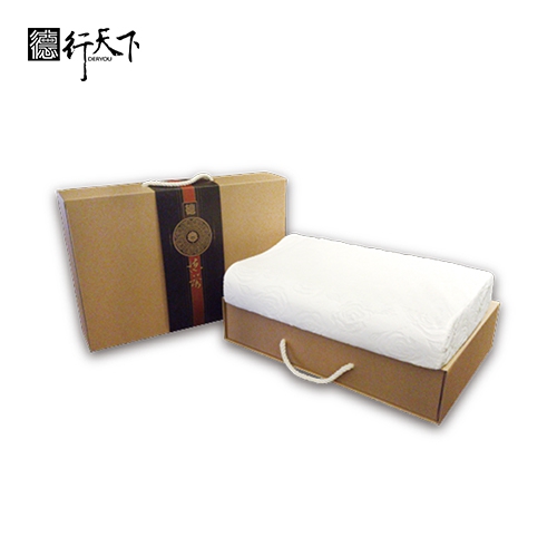
Our team supports comprehensive branding solutions, including logo printing, custom packaging, and product integration support for marketing campaigns. Whether you're launching a new product line or upgrading an existing one, we help your vision come to life with attention to detail and consistent brand presentation.
With fast prototyping services and efficient lead times, GuangXin helps reduce your time-to-market and respond quickly to evolving trends or seasonal demands. From concept to final production, we offer agile support that keeps you ahead of the competition.
Quality Assurance & Certifications
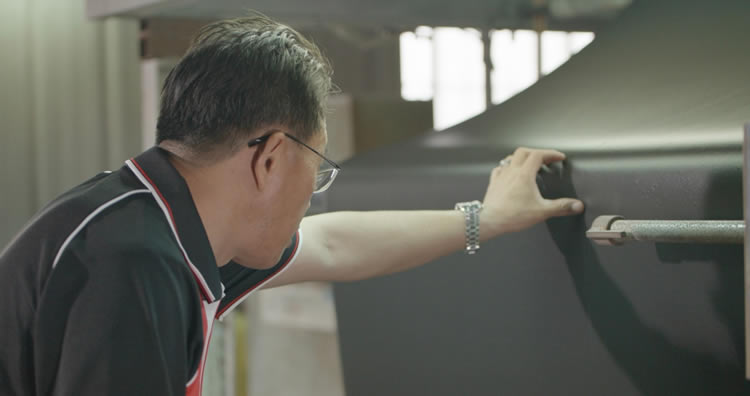
Quality is at the heart of everything we do. GuangXin implements a rigorous quality control system at every stage of production—ensuring that each insole meets the highest standards of consistency, comfort, and durability.
We provide a variety of in-house and third-party testing options, including antibacterial performance, odor control, durability testing, and eco-safety verification, to meet the specific needs of our clients and markets.
Our products are fully compliant with international safety and environmental standards, such as REACH, RoHS, and other applicable export regulations. This ensures seamless entry into global markets while supporting your ESG and product safety commitments.
ESG-Oriented Sustainable Production
At GuangXin Industrial, we are committed to integrating ESG (Environmental, Social, and Governance) values into every step of our manufacturing process. We actively pursue eco-conscious practices by utilizing eco-friendly materials and adopting low-carbon production methods to reduce environmental impact.
To support circular economy goals, we offer recycled and upcycled material options, including innovative applications such as recycled glass and repurposed LCD panel glass. These materials are processed using advanced techniques to retain performance while reducing waste—contributing to a more sustainable supply chain.
We also work closely with our partners to support their ESG compliance and sustainability reporting needs, providing documentation, traceability, and material data upon request. Whether you're aiming to meet corporate sustainability targets or align with global green regulations, GuangXin is your trusted manufacturing ally in building a better, greener future.
Let’s Build Your Next Insole Success Together
Looking for a reliable insole manufacturing partner that understands customization, quality, and flexibility? GuangXin Industrial Co., Ltd. specializes in high-performance insole production, offering tailored solutions for brands across the globe. Whether you're launching a new insole collection or expanding your existing product line, we provide OEM/ODM services built around your unique design and performance goals.
From small-batch custom orders to full-scale mass production, our flexible insole manufacturing capabilities adapt to your business needs. With expertise in PU, latex, and graphene insole materials, we turn ideas into functional, comfortable, and market-ready insoles that deliver value.
Contact us today to discuss your next insole project. Let GuangXin help you create custom insoles that stand out, perform better, and reflect your brand’s commitment to comfort, quality, and sustainability.
🔗 Learn more or get in touch:
🌐 Website: https://www.deryou-tw.com/
📧 Email: shela.a9119@msa.hinet.net
📘 Facebook: facebook.com/deryou.tw
📷 Instagram: instagram.com/deryou.tw
Breathable insole ODM development Vietnam
Are you looking for a trusted and experienced manufacturing partner that can bring your comfort-focused product ideas to life? GuangXin Industrial Co., Ltd. is your ideal OEM/ODM supplier, specializing in insole production, pillow manufacturing, and advanced graphene product design.
With decades of experience in insole OEM/ODM, we provide full-service manufacturing—from PU and latex to cutting-edge graphene-infused insoles—customized to meet your performance, support, and breathability requirements. Our production process is vertically integrated, covering everything from material sourcing and foaming to molding, cutting, and strict quality control.Soft-touch pillow OEM service in Thailand
Beyond insoles, GuangXin also offers pillow OEM/ODM services with a focus on ergonomic comfort and functional innovation. Whether you need memory foam, latex, or smart material integration for neck and sleep support, we deliver tailor-made solutions that reflect your brand’s values.
We are especially proud to lead the way in ESG-driven insole development. Through the use of recycled materials—such as repurposed LCD glass—and low-carbon production processes, we help our partners meet sustainability goals without compromising product quality. Our ESG insole solutions are designed not only for comfort but also for compliance with global environmental standards.High-performance graphene insole OEM factory Taiwan
At GuangXin, we don’t just manufacture products—we create long-term value for your brand. Whether you're developing your first product line or scaling up globally, our flexible production capabilities and collaborative approach will help you go further, faster.Indonesia athletic insole OEM supplier
📩 Contact us today to learn how our insole OEM, pillow ODM, and graphene product design services can elevate your product offering—while aligning with the sustainability expectations of modern consumers.Indonesia OEM/ODM hybrid insole services
A 3-dimensional model of a natural killer cell (purple) with granules (yellow) attaching to a target cell (gray). A pseudo color scale shows differences in packing density of lipids on NK cell membrane, with warmer colors indicating higher density. Credit: Orange lab, CUIMC A newly discovered fat ‘shield’ that prevents natural killer cells from being destroyed by their own deadly biological weapons also allows some cancer cells to evade an immune system attack, a study at Columbia University has found. The findings, which may lead to new treatments for aggressive cancers, were reported on August 3, 2021, in the journal PLoS Biology by scientists in the Department of Pediatrics at Columbia University Vagelos College of Physicians and Surgeons. Natural killer cells are prolific assassins Natural killer cells are our body’s first line of defense against pathogens and cancer cells, always present and ready to strike at a moment’s notice. Natural killer cells are efficient assassins that can eliminate up to six infected or cancer cells each day. The deadly immune cells grab onto their target and blast it with toxic substances—proteins and enzymes—that punch holes in the cell’s membrane. These substances are not especially selective and could easily destroy the natural killer cell’s membrane during the attack. But if these substances are so deadly, how do natural killer cells survive the blast? “I’ve been working on natural killer cells since the early 1990s, and every time I gave a talk about these cells, someone always asked that question,” says study leader and immunology expert Jordan Orange, MD, PhD, the Reuben S. Carpentier Professor of Pediatrics and chair of the Department of Pediatrics at Columbia University Vagelos College of Physicians and Surgeons. “And nobody really knew until now.” Shielded by fat Yu Li, a graduate student working with Orange to understand how natural killer cells work and co-author of the study, thought the answer might lay in the double layer of lipids—a type of fat—that makes up the outer membranes of all cells. Compared with other cells, Li noticed, the membranes of natural killer cells looked more orderly and more densely packed with lipids when viewed under a microscope. “There were a lot of hypotheses about why natural killer cells don’t kill themselves during their attack on other cells, but they all proposed there might be a magic, unknown protein protecting these cells,” Li says. But Li had doubts. “Based on biophysical considerations, I didn’t think a protein would be strong enough to protect the cells. When I looked at the cells, I thought of lipids.” Li put his theory to the test: he exposed the membranes to a compound that weakens the structure of the lipid layer. With less dense and less orderly membranes, the natural killer cells were unprotected from their own toxic blast—and perished along with their targets. Reinforcement arrives before natural killer cells attack To ensure their ability to survive, natural killer cells reinforce their membranes immediately before they launch an attack, Li found. Small granules that contain the deadly substances move to the outer edge of the natural killer cell. As the granule releases its cargo into the space between the killer and target cells, its own unusually dense lipid membrane merges with and reinforces the natural killer cell membrane. “In essence, Li found that the membrane turns into a blast shield,” Orange says. “And the protection comes from the way the membrane’s lipids are arranged. When the lipids are arranged in a more orderly fashion, more lipids can be packed into the membrane. The toxic substances simply can’t find a way into the membrane,” Orange says. Lipid blast shield also protects some cancer cells Natural killer cells are not the only ones to adopt lipid blast shields, Li and Orange also found. At least some cancer cells have adopted the defense to protect themselves during attacks by natural killer cells (and possibly from cytotoxic T cells, another immune cell that uses lipids for protection). Li found that cells from an aggressive breast cancer known to be impervious to natural killer cells fortify their membranes during the attack. The fortification is essential in protecting the cancer cells, Li discovered, because when he added a membrane compound that disrupts lipid packing, the cancer cells were made vulnerable. “We don’t know yet if this is a general mechanism by which cancer cells resist natural killer cells,” Li says. “If it is generalizable, we can start to think of therapies that disrupt the tumor cell membrane and make it more susceptible to attack by the immune system.” Reference: “Degranulation enhances presynaptic membrane packing, which protects NK cells from perforin-mediated autolysis” by Yu Li and Jordan S. Orange, 3 August 2021, PLoS Biology. DOI: 10.1371/journal.pbio.3001328 The study was supported by the National Institutes of Health (R01 AI067946-14).
Using a new method, scientists have discovered that cells lose about 4% of their mass as they enter cell division. They are essentially taking out the trash to give their offspring a fresh start. Cells may use this strategy to clear out toxic byproducts and give their offspring a fresh start. MIT scientists have discovered that before cells start to divide, they do a little cleanup, throwing out molecules that they appear not to need anymore. Using a new method they developed for measuring the dry mass of cells, the researchers found that cells lose about 4 percent of their mass as they enter cell division. The researchers believe that this emptying of trash helps cells to give their offspring a “clean slate,” free of the parent cell’s accumulated junk. “Our hypothesis is that cells might be throwing out things that are building up, toxic components or just things that don’t function properly that you don’t want to have there. It could allow the newborn cells to be born with more functional contents,” says Teemu Miettinen, an MIT research scientist and the lead author of the new study. Scott Manalis, the David H. Koch Professor of Engineering in the departments of Biological Engineering and Mechanical Engineering, and a member of the Koch Institute for Integrative Cancer Research, is the senior author of the paper, which was published on May 10, 2022, in the journal eLife. MIT biological engineering undergraduates Kevin Ly and Alice Lam are also authors of the paper. Measuring Mass Measuring the dry mass of a cell — the weight of its contents not including the water — is commonly done using a microscopy technique called quantitative phase microscopy. This technique can measure cell growth, but it does not reveal information about the molecular content of the dry mass and it is difficult to use with cells that grow in suspension. Manalis’ lab has previously developed a technique for measuring the buoyant mass of cells, which is their mass as they float in a fluid such as water. This method measures buoyant mass by flowing cells through a channel embedded in a vibrating cantilever, which can be done repeatedly to track changes in a particular cell’s mass over many hours or days. MIT researchers have discovered that before cells start to divide, they toss waste products. In this image, the magenta represents DNA, and the green represents a lysosomal marker on the surface of the cells, which is an indicator of lysosomal exocytosis. Credit: Courtesy of the researchers For their new study, the researchers wanted to adapt the technique so that it could be used to calculate the dry mass of cells, as well as the density of the dry mass. About 10 years ago, they had discovered that they could calculate a cell’s dry mass if they first measured the cell in normal water and then in heavy water (which contains deuterium instead of ordinary hydrogen). These two measurements can be used to calculate the cell’s dry mass. However, heavy water is toxic to cells, so they were only able to obtain a single measurement per cell. Last year, Miettinen set out to see if he could design a system in which cells could be measured repeatedly with minimal exposure to heavy water. In the system he came up with, cells are exposed to heavy water very briefly as they flow through microfluidic channels. It takes only one second for a cell to completely exchange its water content, so the researchers could measure the cell’s mass when it was full of heavy water, compare it to the mass in normal water, and then calculate the dry mass. “Our idea was that if we minimize the cells’ exposure to the heavy water, we could engineer the system so that we could repeat this measurement over extended time periods without hurting the cell,” Miettinen says. “That enabled us for the first time to track not just the dry mass of a cell, which is what others do using microscopic methods, but also the density of the dry mass, which informs us of the cell’s biomolecular composition.” The researchers showed that their dry mass measurements qualitatively agreed with previous work using quantitative phase microscopy. And, in addition to providing density of the dry mass, the MIT team’s method enables higher temporal resolution, which proved to be useful for revealing dynamics during mitosis (cell division). Taking Out the Trash In cells undergoing mitosis, the researchers used their new technique to study what happens to cell mass and composition during that process. In a 2019 paper, Miettinen and Manalis found that buoyant mass increases slightly as mitosis begins. However, other studies that used quantitative phase microscopy suggested that cells might retain or lose dry mass early in cell division. In the new study, the MIT team measured three types of cancer cells, which are easier to study because they divide more frequently than healthy cells. To their surprise, the researchers found that the dry mass of cells actually decreases when they enter the cell division cycle. This mass is regained later on, before division is complete. Further experiments revealed that as cells enter mitosis, they ramp up activity of a process called lysosomal exocytosis. Lysosomes are cell organelles that break down or recycle cellular waste products, and exocytosis is the process they use to jettison any molecules that aren’t needed anymore. The researchers also found that the density of the dry mass increases as the cells lose dry mass, leading them to believe that the cells are losing low-density molecules such as lipids or lipoproteins. They hypothesize that cells use this process to clear out toxic molecules before dividing. “What we are seeing is that cells might be trying to throw out damaged components before dividing,” Miettinen says. The researchers speculate that their findings may help explain why neurons, which do not divide, are more likely to accumulate toxic proteins such as Tau or amyloid beta, which are linked to the development of Alzheimer’s disease. The findings could also be relevant to cancer: Cancer cells can expel some chemotherapy drugs using exocytosis, helping them to become resistant to the drugs. In theory, preventing exocytosis from occurring before cell division could help to make cancer cells more susceptible to such drugs. “There are diseases where we might want upregulate exocytosis, for example in neurodegenerative diseases, but then there are diseases like cancer where maybe we want to dial it down,” Miettinen says. “In the future, if we could better understand the molecular mechanism behind this, and find a way to trigger it outside of mitosis or prevent it during mitosis, we could really have a new toggle to use when treating disease.” Reference: “Single-cell monitoring of dry mass and dry mass density reveals exocytosis of cellular dry contents in mitosis” by Teemu P Miettinen, Kevin S Ly, Alice Lam, Scott R Manalis, 10 May 2022, eLife. DOI: 10.7554/eLife.76664 The research was funded by the MIT Center for Cancer Precision Medicine, the Virginia and D.K. Ludwig Fund for Cancer Research, the Cancer Systems Biology Consortium, and the Koch Institute Support (core) Grant from the National Cancer Institute.
Researchers at WashU Medicine collaborated with an international team of doctors and scientists to identify the cause of a rare disorder involving intellectual disability and brain malformations. Brain scans from a patient with this disorder reveal atypical features in white matter (arrows, left) and the cerebellum (arrows, right). Credit: Department of Diagnostic and Interventional Neuroradiology at RWTH Aachen University, Germany Researchers at Washington University have discovered a new genetic disorder affecting protein folding, offering potential new treatment avenues for rare brain conditions. When most people feeling unwell visit a doctor, they expect a clear diagnosis and treatment plan. However, for some 30 million Americans with rare diseases, their symptoms don’t match well-known disease patterns, and they may spend years or even lifetimes seeking a diagnosis. Breakthrough in Understanding a New Genetic Disorder Now, a team of researchers from Washington University School of Medicine in St. Louis and international collaborators has solved the mystery of a child with a rare genetic illness that did not fit any known disease. The team found a link between the child’s neurological symptoms and a genetic change that affects how proteins are properly folded within cells, providing the parents with a molecular diagnosis and identifying an entirely new type of genetic disorder. These findings, recently published in the journal Science, could lead to new treatments for rare brain malformations. Researchers at WashU Medicine modeled the effects of a patient’s genetic change in the tiny roundworm, C. elegans. Their findings, published in Science, contributed to the identification of a new type of rare disorder involving intellectual disability and brain malformations. Credit: Matt Miller Advancements in Genetic Research “Many patients with severe, rare genetic disease remain undiagnosed despite extensive medical evaluation,” said Stephen Pak, PhD, a professor of pediatrics and a co-corresponding author on the study. “Our study has helped a family better understand their child’s illness, preventing further unnecessary clinical evaluations and tests. The findings also have made it possible to identify 22 additional patients with the same or overlapping neurological symptoms and genetic changes that affect protein folding, paving the way for even more diagnoses and, ultimately, potential treatments.” According to Pak, about 10% of patients with suspected genetic disorders have a variant in a gene that has not yet been linked to a disease. His career has been focused on solving such medical mysteries. Pak and author Tim Schedl, PhD, a professor of genetics and a co-director of the model organisms screening center at WashU Medicine, use tiny roundworms called C. elegans to assess whether specific genetic changes found in undiagnosed patients are responsible for their symptoms. With funding from the Eunice Kennedy Shriver National Institute of Child Health and Human Development of the National Institutes of Health (NIH), they and a team of researchers at WashU Medicine have committed to solving more such cases. Methodology and Findings For this study, they teamed up with researchers and doctors from more than a dozen institutions across North America, Europe, India, and China to identify the cause of a cluster of clinical findings in a boy from Germany, and other similar cases. The German patient had an intellectual disability, low muscle tone, and a small brain with abnormal structures. Doctors also found changes to the CCT3 gene, so Pak’s team set out to determine if it could be the cause of the patient’s condition. C. elegans has counterparts to about 50% of human genes, including the CCT3 gene, which is known as cct-3 in roundworms. Weimin Yuan, PhD, a staff scientist in pediatrics and co-first author, found that C. elegans with the patient’s genetic variant moved slower than roundworms with a healthy copy of the gene did, revealing that the genetic change can affect mobility and the nervous system. The affected CCT3 protein is part of the large TRIC/CCT molecular complex whose job is to fold other proteins into their proper shape so they function as they should within cells. The study found that the protein-folding machinery cannot perform without a specific amount of healthy CCT3. “We knew the child has one good and one bad variant gene copy,” Schedl said. “Our studies in C. elegans revealed that the genetic change reduces the activity of the normal protein, decreasing the capacity of the protein-folding machinery, and that for both C. elegans cct-3 and human CCT3, having 50% of activity was insufficient for normal biological function.” The outcome of having reduced protein-folding machinery, they found, was that actin proteins – which help to maintain cell shape and movement –were incorrectly folded and abnormally distributed throughout the cells of C. elegans that carried the patient’s variant. “An understanding of the impact of the genetic change informs the treatment modality,” Schedl added, “because the treatment needed to increase the amount of a normal protein differs from the treatment needed when the protein is poisonous or overactive.” Collaborators from RWTH Aachen University in Germany and Stanford University performed complementary investigations into cct3 variants in zebrafish – which illuminated the effects of the gene on brain development – and in yeast, which clarified its role in protein folding, respectively. Implications for Future Treatments and Diagnoses To determine if there were other patients with this same disorder, researchers mined a freely accessible global database of individuals with intellectual and developmental disabilities. They identified 22 individuals with genetic changes in seven of the eight CCT proteins that form the protein-folding machine. Abnormalities in mobility and actin folding were again seen in roundworms with variants affecting CCT1 and CCT7 proteins, just as the WashU Medicine team observed with dysfunctional CCT3. Together, these patients represent a new type of rare genetic disease involving the protein folding machinery. “This work underscores the importance of using simpler model organisms, like C. elegans, to provide novel insights into human pathobiology,” said co-author Gary Silverman, MD, PhD, the Harriet B. Spoehrer Professor of Pediatrics and head of the Department of Pediatrics. “Our findings can inform clinicians, the scientific community, and patients and families all around the world that changes to the genetic message that are needed to make the eight-protein complex cause disease,” added Pak, who together with Schedl and a team of NIH-funded researchers at WashU Medicine, aim to solve challenging medical mysteries using advanced technologies. “If next week a patient with brain malformations and neurological symptoms is found to have a variant that affects the protein-folding machine, the patient will receive a diagnosis.” Reference: “Brain malformations and seizures by impaired chaperonin function of TRiC” by Florian Kraft, Piere Rodriguez-Aliaga, Weimin Yuan, Lena Franken, Kamil Zajt, Dimah Hasan, Ting-Ting Lee, Elisabetta Flex, Andreas Hentschel, A. Micheil Innes, Bixia Zheng, Dong Sun Julia Suh, Cordula Knopp, Eva Lausberg, Jeremias Krause, Xiaomeng Zhang, Pamela Trapane, Riley Carroll, Martin McClatchey, Andrew E. Fry, Lisa Wang, Sebastian Giesselmann, Hieu Hoang, Dustin Baldridge, Gary A. Silverman, Francesca Clementina Radio, Enrico Bertini, Andrea Ciolfi, Katherine A Blood, Jean-Madeleine de Sainte Agathe, Perrine Charles, Gaber Bergant, Goran Čuturilo, Borut Peterlin, Karin Diderich, Haley Streff, Laurie Robak, Renske Oegema, Ellen van Binsbergen, John Herriges, Carol J. Saunders, Andrea Maier, Stefan Wolking, Yvonne Weber, Hanns Lochmüller, Stefanie Meyer, Alberto Aleman, Kiran Polavarapu, Gael Nicolas, Alice Goldenberg, Lucie Guyant, Kathleen Pope, Katherine N. Hehmeyer, Kristin G. Monaghan, Annegret Quade, Thomas Smol, Roseline Caumes, Sarah Duerinckx, Chantal Depondt, Wim Van Paesschen, Claudine Rieubland, Claudia Poloni, Michel Guipponi, Severine Arcioni, Marije Meuwissen, Anna C. Jansen, Jessica Rosenblum, Tobias B. Haack, Miriam Bertrand, Lea Gerstner, Janine Magg, Olaf Riess, Jörg B. Schulz, Norbert Wagner, Martin Wiesmann, Joachim Weis, Thomas Eggermann, Matthias Begemann, Andreas Roos, Martin Häusler, Tim Schedl, Marco Tartaglia, Juliane Bremer, Stephen C. Pak, Judith Frydman, Miriam Elbracht and Ingo Kurth, 31 October 2024, Science. DOI: 10.1126/science.adp8721 This work was support by the National Institute of Child Health and Human Development of the National Institutes of Health (NIH), grant number R01 HD110556; the NIH, grant numbers GM74074 and GM56433; the Children’s Discovery Institute, St Louis Children’s Hospital Foundation; Italian Ministry of Health, grant numbers RCR-2022-23682289 and PNRR-MR1-2022-12376811; the Canadian Institutes of Health Research (CIHR) for Foundation Grant, grant number FDN-167281; the Transnational Team Grant, grant number ERT-174211; the Network Grant OR2-189333, grant number NMD4C; the Canada Foundation for Innovation, grant number CFI-JELF 38412; the Canada Research Chairs program (Canada Research Chair in Neuromuscular Genomics and Health), grant number 950-232279; the European Commission, grant number 101080249; the Canada Research Coordinating Committee New Frontiers in Research Fund, grant number NFRFG-2022-00033and from the Government of Canada, Canada First Research Excellence Fund (CFREF) for the Brain-Heart Interconnectome, grant number CFREF-2022- 00007; CIHR Postdoctoral fellowship; the German Research Foundation, grant number WO 2385/2-1; the Deutsche Forschungsgemeinschaft (DFG, German Research Foundation), grant numbers WE 1406/16-1, WE 1406/17-1, 418081722, 433158657, 499059538, INST 222/1458-1 FUGG, KU 1587/6-1, KU 1587/9-1, KU 1587/10-1 and KU 1587/11-1; the “Ministerium für Kultur und Wissenschaft des Landes Nordrhein-Westfalen,” grant number PROFILNRW-2020–107-A; “Der Regierende Bürgermeister von Berlin, Senatskanzlei Wissenschaft und Forschung”; postdoctoral fellowship from The Hereditary Disease Foundation (2019-2023); the lonGER consortium; the European Union’s Horizon 2020 research and innovation programme under the EJP RD COFUND-EJP, grant number 825575. The content is solely the responsibility of the authors and does not necessarily represent the views of the NIH.
DVDV1551RTWW78V
Graphene insole manufacturer in Taiwan 》reducing complexity, increasing product valueSmart pillow ODM manufacturing factory Taiwan 》delivering consistent quality from sample to mass productionCushion insole OEM solution China 》the smart choice for brands seeking quality and customization
限會員,要發表迴響,請先登入


