Introduction – Company Background
GuangXin Industrial Co., Ltd. is a specialized manufacturer dedicated to the development and production of high-quality insoles.
With a strong foundation in material science and footwear ergonomics, we serve as a trusted partner for global brands seeking reliable insole solutions that combine comfort, functionality, and design.
With years of experience in insole production and OEM/ODM services, GuangXin has successfully supported a wide range of clients across various industries—including sportswear, health & wellness, orthopedic care, and daily footwear.
From initial prototyping to mass production, we provide comprehensive support tailored to each client’s market and application needs.
At GuangXin, we are committed to quality, innovation, and sustainable development. Every insole we produce reflects our dedication to precision craftsmanship, forward-thinking design, and ESG-driven practices.
By integrating eco-friendly materials, clean production processes, and responsible sourcing, we help our partners meet both market demand and environmental goals.


Core Strengths in Insole Manufacturing
At GuangXin Industrial, our core strength lies in our deep expertise and versatility in insole and pillow manufacturing. We specialize in working with a wide range of materials, including PU (polyurethane), natural latex, and advanced graphene composites, to develop insoles and pillows that meet diverse performance, comfort, and health-support needs.
Whether it's cushioning, support, breathability, or antibacterial function, we tailor material selection to the exact requirements of each project-whether for foot wellness or ergonomic sleep products.
We provide end-to-end manufacturing capabilities under one roof—covering every stage from material sourcing and foaming, to precision molding, lamination, cutting, sewing, and strict quality control. This full-process control not only ensures product consistency and durability, but also allows for faster lead times and better customization flexibility.
With our flexible production capacity, we accommodate both small batch custom orders and high-volume mass production with equal efficiency. Whether you're a startup launching your first insole or pillow line, or a global brand scaling up to meet market demand, GuangXin is equipped to deliver reliable OEM/ODM solutions that grow with your business.
Customization & OEM/ODM Flexibility
GuangXin offers exceptional flexibility in customization and OEM/ODM services, empowering our partners to create insole products that truly align with their brand identity and target market. We develop insoles tailored to specific foot shapes, end-user needs, and regional market preferences, ensuring optimal fit and functionality.
Our team supports comprehensive branding solutions, including logo printing, custom packaging, and product integration support for marketing campaigns. Whether you're launching a new product line or upgrading an existing one, we help your vision come to life with attention to detail and consistent brand presentation.
With fast prototyping services and efficient lead times, GuangXin helps reduce your time-to-market and respond quickly to evolving trends or seasonal demands. From concept to final production, we offer agile support that keeps you ahead of the competition.
Quality Assurance & Certifications
Quality is at the heart of everything we do. GuangXin implements a rigorous quality control system at every stage of production—ensuring that each insole meets the highest standards of consistency, comfort, and durability.
We provide a variety of in-house and third-party testing options, including antibacterial performance, odor control, durability testing, and eco-safety verification, to meet the specific needs of our clients and markets.
Our products are fully compliant with international safety and environmental standards, such as REACH, RoHS, and other applicable export regulations. This ensures seamless entry into global markets while supporting your ESG and product safety commitments.
ESG-Oriented Sustainable Production
At GuangXin Industrial, we are committed to integrating ESG (Environmental, Social, and Governance) values into every step of our manufacturing process. We actively pursue eco-conscious practices by utilizing eco-friendly materials and adopting low-carbon production methods to reduce environmental impact.
To support circular economy goals, we offer recycled and upcycled material options, including innovative applications such as recycled glass and repurposed LCD panel glass. These materials are processed using advanced techniques to retain performance while reducing waste—contributing to a more sustainable supply chain.
We also work closely with our partners to support their ESG compliance and sustainability reporting needs, providing documentation, traceability, and material data upon request. Whether you're aiming to meet corporate sustainability targets or align with global green regulations, GuangXin is your trusted manufacturing ally in building a better, greener future.
Let’s Build Your Next Insole Success Together
Looking for a reliable insole manufacturing partner that understands customization, quality, and flexibility? GuangXin Industrial Co., Ltd. specializes in high-performance insole production, offering tailored solutions for brands across the globe. Whether you're launching a new insole collection or expanding your existing product line, we provide OEM/ODM services built around your unique design and performance goals.
From small-batch custom orders to full-scale mass production, our flexible insole manufacturing capabilities adapt to your business needs. With expertise in PU, latex, and graphene insole materials, we turn ideas into functional, comfortable, and market-ready insoles that deliver value.
Contact us today to discuss your next insole project. Let GuangXin help you create custom insoles that stand out, perform better, and reflect your brand’s commitment to comfort, quality, and sustainability.
🔗 Learn more or get in touch:
🌐 Website: https://www.deryou-tw.com/
📧 Email: shela.a9119@msa.hinet.net
📘 Facebook: facebook.com/deryou.tw
📷 Instagram: instagram.com/deryou.tw
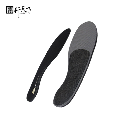
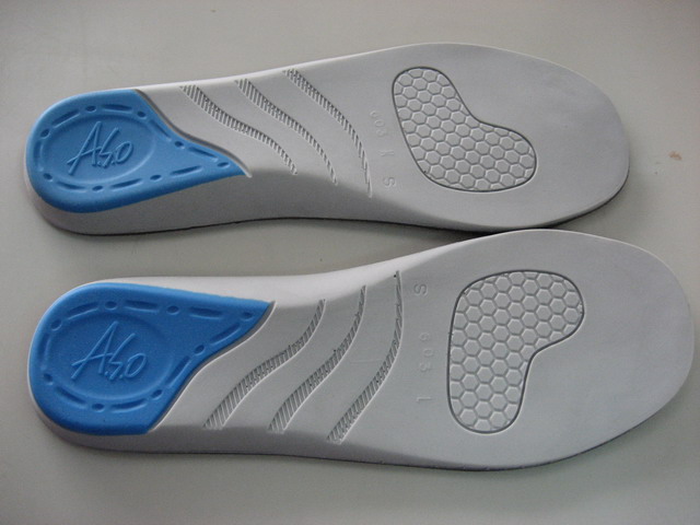
Customized sports insole ODM Indonesia
Are you looking for a trusted and experienced manufacturing partner that can bring your comfort-focused product ideas to life? GuangXin Industrial Co., Ltd. is your ideal OEM/ODM supplier, specializing in insole production, pillow manufacturing, and advanced graphene product design.
With decades of experience in insole OEM/ODM, we provide full-service manufacturing—from PU and latex to cutting-edge graphene-infused insoles—customized to meet your performance, support, and breathability requirements. Our production process is vertically integrated, covering everything from material sourcing and foaming to molding, cutting, and strict quality control.Indonesia custom product OEM/ODM services
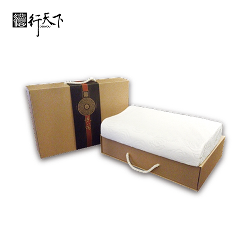
Beyond insoles, GuangXin also offers pillow OEM/ODM services with a focus on ergonomic comfort and functional innovation. Whether you need memory foam, latex, or smart material integration for neck and sleep support, we deliver tailor-made solutions that reflect your brand’s values.
We are especially proud to lead the way in ESG-driven insole development. Through the use of recycled materials—such as repurposed LCD glass—and low-carbon production processes, we help our partners meet sustainability goals without compromising product quality. Our ESG insole solutions are designed not only for comfort but also for compliance with global environmental standards.Custom foam pillow OEM production factory in Taiwan
At GuangXin, we don’t just manufacture products—we create long-term value for your brand. Whether you're developing your first product line or scaling up globally, our flexible production capabilities and collaborative approach will help you go further, faster.Vietnam insole ODM design and production
📩 Contact us today to learn how our insole OEM, pillow ODM, and graphene product design services can elevate your product offering—while aligning with the sustainability expectations of modern consumers.Graphene insole OEM factory Vietnam
Researchers developed a new epigenome editing platform that allows precise manipulation of chromatin marks, revealing their direct impact on gene expression and challenging previous understanding of gene regulation mechanisms. A study from the Hackett group at EMBL Rome led to the development of a powerful epigenetic editing technology, which unlocks the ability to precisely program chromatin modifications. Understanding how genes are regulated at the molecular level is a central challenge in modern biology. This complex mechanism is mainly driven by the interaction between proteins called transcription factors, DNA regulatory regions, and epigenetic modifications – chemical alterations that change chromatin structure. The set of epigenetic modifications of a cell’s genome is referred to as the epigenome. Advancements in Epigenome Editing In a study published today (May 9) in Nature Genetics, scientists from the Hackett Group at European Molecular Biology Laboratory (EMBL) Rome have developed a modular epigenome editing platform – a system to program epigenetic modifications at any location in the genome. The system allows scientists to study the impact of each chromatin modification on transcription, the mechanism by which genes are copied into mRNA to drive protein synthesis. Chromatin modifications are thought to contribute to the regulation of key biological processes such as development, response to environmental signals, and disease. Creative depiction of the epigenetic editing toolkit: each building represents the epigenetic state of a single gene (dark windows are silenced genes, lit up windows are active genes). The crane illustrates the epigenetic editing system which enables de novo deposition of chromatin marks on any genomic location. Marzia Munafò To understand the effects of specific chromatin marks on gene regulation, previous studies have mapped their distribution in the genomes of healthy and diseased cell types. By combining this data with gene expression analysis and the known effects of perturbing specific genes, scientists have ascribed functions to such chromatin marks. However, the causal relationship between chromatin marks and gene regulation has proved difficult to determine. The challenge lies in dissecting the individual contributions of the many complex factors involved in such regulation – chromatin marks, transcription factors, and regulatory DNA sequences. Breakthrough in Epigenome Editing Technology Scientists from the Hackett Group developed a modular epigenome editing system to precisely program nine biologically important chromatin marks at any desired region in the genome. The system is based on CRISPR – a widely used genome editing technology that allows researchers to make alterations in specific DNA locations with high precision and accuracy. Such precise perturbations enabled them to carefully dissect cause-and-consequence relationships between chromatin marks and their biological effects. The scientists also designed and employed a ‘reporter system’, which allowed them to measure changes in gene expression at single-cell level and to understand how changes in the DNA sequence influence the impact of each chromatin mark. Their results reveal the causal roles of a range of important chromatin marks in gene regulation. Key Findings and Future Directions For example, the researchers found a new role for H3K4me3, a chromatin mark that was previously believed to be a result of transcription. They observed that H3K4me3 can actually increase transcription by itself if artificially added to specific DNA locations. “This was an extremely exciting and unexpected result that went against all our expectations,” said Cristina Policarpi, postdoc in the Hackett Group and leading scientist of the study. “Our data point towards a complex regulatory network, in which multiple governing factors interact to modulate the levels of gene expression in a given cell. These factors include the pre-existing structure of the chromatin, the underlying DNA sequence, and the location in the genome.” Potential Applications and Future Research Hackett and colleagues are currently exploring avenues to leverage this technology through a promising start-up venture. The next step will be to confirm and expand these conclusions by targeting genes across different cell types and at scale. How chromatin marks influence transcription across the diversity of genes and downstream mechanisms, also remains to be clarified. “Our modular epigenetic editing toolkit constitutes a new experimental approach to dissect the reciprocal relationships between the genome and epigenome,” said Jamie Hackett, Group Leader at EMBL Rome. “The system could be used in the future to more precisely understand the importance of epigenomic changes in influencing gene activity during development and in human disease. On the other hand, the technology also unlocks the ability to program desired gene expression levels in a highly tunable manner. This is an exciting avenue for precision health applications and may prove useful in disease settings.” Reference: “Systematic epigenome editing captures the context-dependent instructive function of chromatin modifications” by Cristina Policarpi, Marzia Munafò, Stylianos Tsagkris, Valentina Carlini and Jamie A. Hackett, 9 May 2024, Nature Genetics. DOI: 10.1038/s41588-024-01706-w
Illustration of the interacting thick and thin filaments in the cardiac sarcomere based on structural cryo electron-tomography data. Credit: MPI of Molecular Physiology Scientists captured the first true-to-life 3D image of the thick filament in mammalian heart muscle. Atrial fibrillation, heart failure, and stroke are among the severe health conditions that can stem from hypertrophic cardiomyopathy, a critical factor in sudden cardiac death among individuals under 35 years old. “The heart muscle is a central engine of the human body. Of course, it is easier to fix a broken engine, if you know how it is built and how it functions,” says Stefan Raunser. “At the beginning of our muscle research, we have successfully visualized the structure of the essential muscle building blocks and how they interact using electron cryo-microscopy.” “However, these were static images of proteins taken out of the living cell. They only tell us little about how the highly variable, dynamic interplay of muscle components moves the muscle in its native environment,” says Raunser. Through thick and thin Skeletal and heart muscles contract upon the interaction of two types of parallel protein filaments in the sarcomere: thin and thick. The sarcomere is subdivided in several regions, called zones and bands, in which these filaments are arranged in different ways. The thin filament consists of F-actin, troponin, tropomyosin, and nebulin. The thick filament is formed of myosin, titin, and myosin-binding protein C (MyBP-C). The latter can form links between the filaments, whereas myosin, the so-called motor protein interacts with the thin filament to generate force and muscle contraction. Thick filament structure in the relaxed cardiac sarcomere. The upper image shows a tomographic slice of a cardiac sarcomere. Thin filaments are marked with green and thick filaments with a purple arrow. The middle image shows the reconstructed thick (purple) and thin (green) filaments. The lower image shows the structure of the thin filament spanning across several sarcomere regions. The scale bar shows 50 nm. Credit:MPI of Molecular Physiology Alterations in the thick filament proteins are associated with muscle diseases. A detailed picture of the thick filament would be of immense importance for developing therapeutical strategies to cure these diseases, but has been missing so far. Milestones in muscle research “If you want to fully understand how the muscle works on the molecular level, you need to picture its components in their natural environment – one of the biggest challenges in biological research nowadays that cannot be tackled by traditional experimental approaches,” says Raunser. To overcome this obstacle his team developed an electron cryo-tomography workflow specifically tailored to the investigation of muscle samples: The scientists flash-freeze mammalian heart muscle samples, produced by the Gautel group in London, at a very low temperature (- 175 °C). 3D structure of the sarcomere showing thick (purple) and thin (green) filaments. Credit: MPI of Molecular Physiology This preserves their hydration and fine structure and thus their native state. A focused ion beam (FIB milling) is then applied to thin out the samples to an ideal thickness of around 100 nanometers for the transmission electron microscope, which acquires multiple images as the sample is tilted along an axis. Finally, computational methods reconstruct a three-dimensional picture at high resolution. In recent years, Raunser’s group successfully applied the customized workflow, resulting in two recent groundbreaking publications: They produced the first high-resolution images of the sarcomere and of a so far nebulous muscle protein called nebulin. Both studies provide unprecedented insights into the 3D organization of muscle proteins in the sarcomere, e. g. how myosin binds to actin to control muscle contraction and how nebulin binds to actin to stabilize it and to determine its length. Completing the painting In their current study, the scientists produced the first high-resolution image of the cardiac thick filament spanning across several regions in the sarcomere. “With 500 nm length this makes for the longest and biggest structure ever resolved by cryo-ET,” says Davide Tamborrini from the MPI Dortmund, first-author of the study. Even more impressive are the newly gained insights into the thick filament’s molecular organization and thus into its function. The arrangement of the myosin molecules depends on their position in the filament. The scientists suspect, that this allows the thick filament to sense and process numerous muscle-regulating signals and thus to regulate the strength of muscle contraction depending on the sarcomere region. They also revealed how titin chains run along the filament. Titin chains intertwine with myosin, acting as a scaffold for its assembly and probably orchestrating a length-depending activation of the sarcomere. “Our aim is to paint a complete picture of the sarcomere one day. The image of the thick filament in this study is ‘only’ a snapshot in the relaxed state of the muscle. To fully understand how the sarcomere functions and how it is regulated, we want to analyze it in different states e. g. during contraction,” says Raunser. Comparison with samples from patients with muscle disease will ultimately contribute to a better understanding of diseases like hypertrophic cardiomyopathy and to the development of innovative therapies. Reference: “Structure of the native myosin filament in the relaxed cardiac sarcomere” by Davide Tamborrini, Zhexin Wang, Thorsten Wagner, Sebastian Tacke, Markus Stabrin, Michael Grange, Ay Lin Kho, Martin Rees, Pauline Bennett, Mathias Gautel and Stefan Raunser, 1 November 2023, Nature. DOI: 10.1038/s41586-023-06690-5
Light microscope images of euglenoid cysts from the Triassic-Jurassic boundary (approx. 200 million years old) in the Schandelah-1 core in Germany (left) and from Triassic sediments in Winterswijk, the Netherlands (right). The specimens are between 20 and 30 micrometers in diameter. Credit: Bas van de Schootbrugge, Utrecht University A 400-million-year evolutionary history. Euglenoids, a unique group of single-celled protists, occupy a mysterious niche, being neither fully plant nor animal. Unlike plants that solely rely on photosynthesis, or animals that consume, euglenoids embrace both modes. They navigate the dim waters of shallow freshwater ponds, propelled by their elongated flagella, consuming organic matter while simultaneously harnessing their chloroplasts to transform CO2, water, and light into sugars. This dual nature positions euglenoids near the foundational root of the eukaryotic branch on the life tree, encompassing plants, fungi, and animals. Despite their ancient origins, believed to date back over 1 billion years, euglenoids have left behind a remarkably sparse fossil record. In a new study published in the journal Review of Palaeobotany and Palynology, a team of Dutch, American, British, German, and Australian scientists shed new light on a group of “problematic” microfossils that have remained a mystery for nearly a century. By comparing microscopic fossil cysts in 200-million-year-old pond sediments from cores drilled in Germany and the Netherlands to much older Paleozoic, and much younger remains in Holocene lakes in Greece, and finally to living protists in a pond in Australia, the researchers establish a 400-million-year evolutionary history of the euglenoids. Transmission Electron Microscope (TEM) image of the wall structure of a Holocene euglenoid cyst from Lake Vouliagmeni, Greece. Credit: Wilson Taylor, University of Wisconsin – Eau Claire What’s in a name? In 2012, Bas van de Schootbrugge, then at the Goethe University in Frankfurt am Main, and Paul Strother from Boston College, while working on a variety of problematic microfossils in sediments from around the Triassic-Jurassic boundary, realized that the circular striated cysts they were seeing, could be in fact euglenoid cysts. “We had this amazing drill core material at our disposal that contained many unidentified microfossils, including some of the oldest butterfly remains that we published on in 2018,” said Bas van de Schootbrugge, now at Utrecht University. Paul Strother continued: “Some of the microfossils we encountered showed a canny similarity to cysts of Euglena, a modern representative that had been described by Slovakian colleagues. The problem was, there was only one publication in the world making this claim.” Video stills from encysting Euglena from New South Wales, Australia. Credit: Fabian Weston, Protist Lab Films, Galston Even more unsettling: after an extensive literature review, van de Schootbrugge and Strother realized that the same type of microfossil had been given many different names. Scientists working on Quaternary and Holocene time slices used Concentricystes, referring to a possible algal cyst with concentric ribs. But Mesozoic workers used Pseudoschizaea, originally thinking it could have been a fern spore. Even older fossils from the Permian, Devonian, Silurian, and Ordovician were known as Circulisporites and Chomotriletes. Transmission electron microscopy After the authors had disentangled the taxonomic confusion, compiling in the process nearly 500 literature sources related to any of the four taxa, more advanced microscope techniques were needed to establish the ultrastructure of the cysts with the help of transmission electron microscopy (TEM). This required picking of single specimens, embedding, and micro-tome slicing by University of Wisconsin-Eau-Claire co-author Wilson Taylor. Because the specimens in the Triassic-Jurassic cores were mostly damaged, the team turned to palynologist Andreas Koutsodendris at Heidelberg University (Germany), who had access to Holocene and Pliocene core samples containing abundant well-preserved specimens. Light microscope images of euglenoid cysts from Holocene to recent Lake Vouliagmeni in Greece. Specimens are between 20 and 30 micrometers in diameter. Note the fingerprint-like patterning that is a shared characteristic of all fossil forms. Credit: Andreas Koutsodendris, Heidelberg University Andreas Koutsodendris said: “I am encountering these cysts regularly in cores drilled in lakes, for example in Lake Vouliagmeni in Greece that we studied here, but their biological affinity has never been cleared. In fact, the cysts are commonly figured in publications by colleagues, but no one was able to really put a finger on it.” Wilson Taylor continued: “We were much surprised by the ultrastructure of the cysts. The structure of the wall does not resemble anything that is known. The ribs are not ornaments, like in pollen and spores, but part of the wall structure,” said Wilson Taylor. “The layered structure of the walls is also clearly different from many other fresh-water green algae,” Taylor continued. Nagging uncertainty While the TEM analysis initially added more mystery, the results did align with a study published in 2021 by another group of colleagues that looked at the ultrastructure of Pseudoschizaea. At least it was possible to show that Holocene and Pliocene Concentricystes and Jurassic Pseudoschizaea are in fact the same. But there remained one nagging uncertainty and that was the lack of any cysts produced by living euglenoids. Wilson Taylor: “We did contact several biologists working on living euglenoids, but no one had been able to make euglenoids encyst in a lab setting, allowing for extraction and TEM analyses of the cysts”. Microscopic life down under Enter Fabian Weston. By chance, Strother and van de Schootbrugge stumbled across superb video material posted on YouTube by microscopy enthusiast Fabian Weston from Sydney, Australia. In 2020 Fabian Weston had put a drop of water sampled from a nearby pond in New South Wales on a microscope slide, and using his advanced set-up at The Protist Lab filmed Euglena as it gracefully moved in and out of focus. For reasons that remain poorly understood but could be related to the drying out of the water under the coverslip, Euglena is then seen to ball up and form a thick wall with ribs that is akin to the cysts found throughout the fossil record. “Unwittingly, Fabian provided a key piece of evidence. He is probably the only person on the planet to have witnessed Euglena encyst under a microscope,” Strother said. Significance and next steps Based on all the available pieces of the puzzle, the authors thus link euglenoids from a pond in Australia to fossil cysts that are more than 400 million years old, establishing a deep time record of the euglenoids. “This opens the door for recognizing even older examples, for example from Precambrian records that go back to the very root of the eukaryotic tree of life,” Strother said. “Now that we know which organisms produced those cysts, we can also use them for paleo-environmental interpretations. Their abundance around two of the largest mass-extinction events of the past 600 million years is a tell-tale sign of some major upheavals on the continents related to increased precipitation under extreme greenhouse climate conditions.” Van de Schootbrugge concluded: “Perhaps related to their capabilities to encyst, these organisms have endured and survived every major extinction on the planet. Unlike the behemoths that were done in by volcanoes and asteroids, these tiny creatures have weathered it all.” Extending their research, the team intends to travel to Australia in the near future to scour for preserved Euglena cysts in pond and lake sediments in New South Wales. Reference: “Recognition of an extended record of euglenoid cysts: Implications for the end-Triassic mass extinction” by Bas van de Schootbrugge, Andreas Koutsodendris, Wilson Taylor, Fabian Weston, Charles Wellman and Paul K. Strother, 21 December 2023, Review of Palaeobotany and Palynology. DOI: 10.1016/j.revpalbo.2023.105043
DVDV1551RTWW78V
China high-end foam product OEM/ODM 》where innovation meets ergonomic comfort and market demandSoft-touch pillow OEM service in Indonesia 》simplifying complex ideas into market-ready productsTaiwan graphene sports insole ODM 》supporting your ESG goals through sustainable production
下一則: Smart pillow ODM manufacturing factory Taiwan 》del
限會員,要發表迴響,請先登入


