Introduction – Company Background
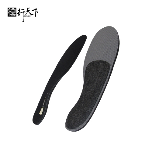
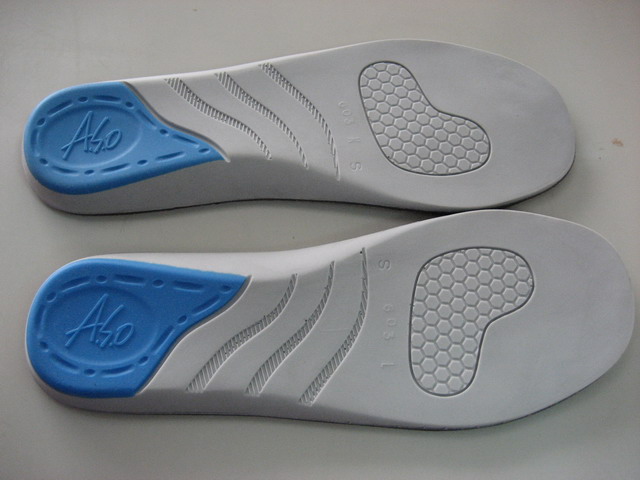
GuangXin Industrial Co., Ltd. is a specialized manufacturer dedicated to the development and production of high-quality insoles.
With a strong foundation in material science and footwear ergonomics, we serve as a trusted partner for global brands seeking reliable insole solutions that combine comfort, functionality, and design.
With years of experience in insole production and OEM/ODM services, GuangXin has successfully supported a wide range of clients across various industries—including sportswear, health & wellness, orthopedic care, and daily footwear.
From initial prototyping to mass production, we provide comprehensive support tailored to each client’s market and application needs.
At GuangXin, we are committed to quality, innovation, and sustainable development. Every insole we produce reflects our dedication to precision craftsmanship, forward-thinking design, and ESG-driven practices.
By integrating eco-friendly materials, clean production processes, and responsible sourcing, we help our partners meet both market demand and environmental goals.
Core Strengths in Insole Manufacturing
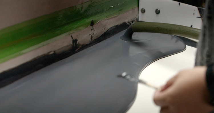
At GuangXin Industrial, our core strength lies in our deep expertise and versatility in insole and pillow manufacturing. We specialize in working with a wide range of materials, including PU (polyurethane), natural latex, and advanced graphene composites, to develop insoles and pillows that meet diverse performance, comfort, and health-support needs.
Whether it's cushioning, support, breathability, or antibacterial function, we tailor material selection to the exact requirements of each project-whether for foot wellness or ergonomic sleep products.
We provide end-to-end manufacturing capabilities under one roof—covering every stage from material sourcing and foaming, to precision molding, lamination, cutting, sewing, and strict quality control. This full-process control not only ensures product consistency and durability, but also allows for faster lead times and better customization flexibility.
With our flexible production capacity, we accommodate both small batch custom orders and high-volume mass production with equal efficiency. Whether you're a startup launching your first insole or pillow line, or a global brand scaling up to meet market demand, GuangXin is equipped to deliver reliable OEM/ODM solutions that grow with your business.
Customization & OEM/ODM Flexibility
GuangXin offers exceptional flexibility in customization and OEM/ODM services, empowering our partners to create insole products that truly align with their brand identity and target market. We develop insoles tailored to specific foot shapes, end-user needs, and regional market preferences, ensuring optimal fit and functionality.
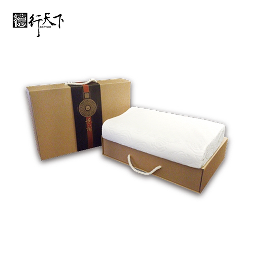
Our team supports comprehensive branding solutions, including logo printing, custom packaging, and product integration support for marketing campaigns. Whether you're launching a new product line or upgrading an existing one, we help your vision come to life with attention to detail and consistent brand presentation.
With fast prototyping services and efficient lead times, GuangXin helps reduce your time-to-market and respond quickly to evolving trends or seasonal demands. From concept to final production, we offer agile support that keeps you ahead of the competition.
Quality Assurance & Certifications
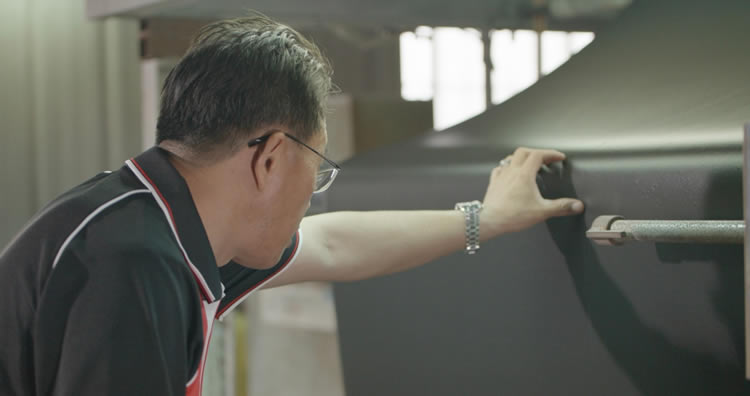
Quality is at the heart of everything we do. GuangXin implements a rigorous quality control system at every stage of production—ensuring that each insole meets the highest standards of consistency, comfort, and durability.
We provide a variety of in-house and third-party testing options, including antibacterial performance, odor control, durability testing, and eco-safety verification, to meet the specific needs of our clients and markets.
Our products are fully compliant with international safety and environmental standards, such as REACH, RoHS, and other applicable export regulations. This ensures seamless entry into global markets while supporting your ESG and product safety commitments.
ESG-Oriented Sustainable Production
At GuangXin Industrial, we are committed to integrating ESG (Environmental, Social, and Governance) values into every step of our manufacturing process. We actively pursue eco-conscious practices by utilizing eco-friendly materials and adopting low-carbon production methods to reduce environmental impact.
To support circular economy goals, we offer recycled and upcycled material options, including innovative applications such as recycled glass and repurposed LCD panel glass. These materials are processed using advanced techniques to retain performance while reducing waste—contributing to a more sustainable supply chain.
We also work closely with our partners to support their ESG compliance and sustainability reporting needs, providing documentation, traceability, and material data upon request. Whether you're aiming to meet corporate sustainability targets or align with global green regulations, GuangXin is your trusted manufacturing ally in building a better, greener future.
Let’s Build Your Next Insole Success Together
Looking for a reliable insole manufacturing partner that understands customization, quality, and flexibility? GuangXin Industrial Co., Ltd. specializes in high-performance insole production, offering tailored solutions for brands across the globe. Whether you're launching a new insole collection or expanding your existing product line, we provide OEM/ODM services built around your unique design and performance goals.
From small-batch custom orders to full-scale mass production, our flexible insole manufacturing capabilities adapt to your business needs. With expertise in PU, latex, and graphene insole materials, we turn ideas into functional, comfortable, and market-ready insoles that deliver value.
Contact us today to discuss your next insole project. Let GuangXin help you create custom insoles that stand out, perform better, and reflect your brand’s commitment to comfort, quality, and sustainability.
🔗 Learn more or get in touch:
🌐 Website: https://www.deryou-tw.com/
📧 Email: shela.a9119@msa.hinet.net
📘 Facebook: facebook.com/deryou.tw
📷 Instagram: instagram.com/deryou.tw
Indonesia custom neck pillow ODM
Are you looking for a trusted and experienced manufacturing partner that can bring your comfort-focused product ideas to life? GuangXin Industrial Co., Ltd. is your ideal OEM/ODM supplier, specializing in insole production, pillow manufacturing, and advanced graphene product design.
With decades of experience in insole OEM/ODM, we provide full-service manufacturing—from PU and latex to cutting-edge graphene-infused insoles—customized to meet your performance, support, and breathability requirements. Our production process is vertically integrated, covering everything from material sourcing and foaming to molding, cutting, and strict quality control.Indonesia graphene sports insole ODM
Beyond insoles, GuangXin also offers pillow OEM/ODM services with a focus on ergonomic comfort and functional innovation. Whether you need memory foam, latex, or smart material integration for neck and sleep support, we deliver tailor-made solutions that reflect your brand’s values.
We are especially proud to lead the way in ESG-driven insole development. Through the use of recycled materials—such as repurposed LCD glass—and low-carbon production processes, we help our partners meet sustainability goals without compromising product quality. Our ESG insole solutions are designed not only for comfort but also for compliance with global environmental standards.Taiwan custom product OEM/ODM manufacturing factory
At GuangXin, we don’t just manufacture products—we create long-term value for your brand. Whether you're developing your first product line or scaling up globally, our flexible production capabilities and collaborative approach will help you go further, faster.China neck support pillow OEM
📩 Contact us today to learn how our insole OEM, pillow ODM, and graphene product design services can elevate your product offering—while aligning with the sustainability expectations of modern consumers.ODM service for ergonomic pillows Taiwan
Scientists have made a major advance toward understanding the molecular mechanisms that are involved in the creation of spatial maps in the brain. The Fos gene plays a key role in forming stable brain maps for navigation, linking molecular processes to memory and behavior. Research in mice illuminates the molecular mechanisms that underlie spatial mapping in the brain Researchers found that a gene called Fos plays a key role in helping the brain use specialized navigation cells to form and maintain spatial maps The findings bring us one step closer to a complete understanding of how the brain creates memories of spatial maps for navigation Anytime we venture into a new location, our brain’s built-in GPS immediately activates and begins to form a spatial map of our surroundings. Over a period of days and even weeks, this map may be solidified as a memory that we can recall to help us navigate more easily whenever we return to that particular place. Just how the brain forms these spatial maps is astoundingly complex. It is a process that involves an intricate molecular interplay across genes, proteins, and neural circuits to shape behavior. Perhaps unsurprisingly given this immense complexity, the precise steps of this multiplayer interaction have eluded neurobiologists. Now, scientists have made a major advance toward understanding the molecular mechanisms that are involved in the creation of spatial maps in the brain. The researchers worked through a multilab collaboration within the Blavatnik Institute at Harvard Medical School. The new study, conducted in mice and published today (August 24, 2022) in the journal Nature, establishes that a gene called Fos is a key player in spatial mapping, helping the brain use specialized navigation cells to form and maintain stable representations of the environment. “This research connects across the different levels of understanding to make a pretty direct link between molecules and the function of circuits for behavior and memory,” said Christopher Harvey, associate professor of neurobiology at HMS and senior author of the study. “Here we can understand what’s actually underlying the formation and stability of spatial maps.” If the findings translate into humans, they will provide crucial new information about how our brains construct spatial maps. Eventually, this knowledge could help researchers better understand what happens when this process breaks down, as it often does as a result of brain injury or neurodegeneration. The Role of the Hippocampus in Navigation and Memory Lying deep in the brain’s temporal lobe, the hippocampus plays an essential role in learning, memory, and navigation for many species, including mice and humans. Scientists have long known that for navigation, the hippocampus contains specialized neurons called place cells that selectively become active when an animal is at different locations in space. By turning on and off as an animal moves through its environment, place cells essentially construct a map of the surrounding area that can be incorporated into a memory. “My lab has studied spatial navigation for years, including how place cells form a map of the environment and form spatial memories,” Harvey said, and yet “the molecular mechanisms that underlie those processes have been difficult to study in the behaving animal.” To study the molecular cascade involved in this mapping process, Harvey and first author Noah Pettit teamed up with co-senior author Michael Greenberg and author Lynn Yap. Pettit is a research fellow in neurobiology in the Harvey lab, Greenberg is the Nathan Marsh Pusey Professor of Neurobiology at HMS, and Yap is a graduate of the Harvard PhD Program in Neuroscience who did her doctoral work in the Greenberg lab. Fos Expression and Its Link to Place Cells Greenberg’s lab studies the Fos gene, which codes for a transcription factor protein that regulates the expression of other genes. In previous research, Greenberg and his colleagues showed that Fos is expressed minutes after a neuron is activated, making it a useful marker for neural activity in the brain. They also demonstrated that Fos acts as a mediator for different types of neural plasticity, including navigation and memory formation. However, the relationship between Fos and place cells in the hippocampus was not known. The team wondered whether Fos could be involved in how mice form spatial maps as they navigate their environment. To find out, the investigators used a technique developed in Harvey’s lab that places mice in a virtual reality maze: A mouse runs on a ball as it looks at a large, surround screen that displays a spatial navigation task such as solving a maze to find a reward. As the mouse jogs on the ball and performs the task, researchers record neural activity and changes in Fos expression in the hippocampus. In what Greenberg called “a technical tour de force,” Pettit led a series of complicated experiments to unravel the connection between Fos and place cells. The researchers found that in the hours after a mouse performed a navigation task, neurons with high Fos expression were more likely to form accurate place fields — clusters of place cells that signal spatial position — than those with low Fos expression. Moreover, neurons with high Fos expression had place fields that were more reliable over time in indicating spatial position as the mouse repeated the task on subsequent days. “This tells us that on a moment-to-moment basis as the mouse is navigating, the neurons that induce Fos have very robust information about the mouse’s spatial position, which is the key variable needed to solve and remember the task,” Pettit explained. When the team knocked out Fos in a subset of neurons within the hippocampus, they observed that those cells had less accurate spatial maps of the environment than nearby neurons with normal Fos expression. Also, the maps in cells lacking Fos were less stable across days, and thus, were less reliable as memories of the environment. Fos’s Role in Maintaining Stable Spatial Maps “Fos seems to be important for maintaining the stability and accuracy of place cells, and representing a spatial map in the brain over time,” Greenberg said. “There have been a lot of studies on Fos and there have been a lot of studies on place cells, but this is one of the first papers that directly connects the two,” Harvey added, “It opens a lot of exciting new directions for investigating these mechanisms.” For instance, Greenberg would like to delve into the specific molecules and cells that are involved as Fos helps the brain form and maintain stable spatial maps over time. He also wants to understand the different roles Fos may play as spatial map memories are transferred from the hippocampus to other brain regions. In a similar vein, Harvey is interested in whether Fos is part of the process by which spatial map memories are solidified during sleep. Although the study was done in mice, the scientists noted that much of the system is conserved across species, including humans. If the findings can be confirmed in humans, they could help scientists understand how our brains form spatial maps and what happens when we lose this ability due to injury or disease. Beyond the science, the researchers emphasized that the research represents an unusual partnership between a laboratory that studies cellular and molecular mechanisms and one that focuses on animal behavior and neural circuits. “Our two laboratories are about as far from each other in terms of what we do as any in the department, but we’ve come together to study how molecules interact with neural circuits that control learning, memory, and behavior,” Greenberg said. “This was a natural and exciting collaboration to learn that Fos plays a role in spatial memories and spatial navigation,” Harvey agreed. “It’s hard to be an expert in all these different levels of neurobiology, but by working together, the two labs have been able to bridge the gap.” Reference: “Fos ensembles encode and shape stable spatial maps in the hippocampus” by Noah L. Pettit, Ee-Lynn Yap, Michael E. Greenberg and Christopher D. Harvey, 24 August 2022, Nature. DOI: 10.1038/s41586-022-05113-1 Funding was provided by the National Institutes of Health (grants DP1 MH125776, R01 NS089521, R01 NS028829), Stuart H.Q. & Victoria Quan Fellowship, HMS Department of Neurobiology graduate fellowship, and Harvard Aramont Fellowship Fund for Emerging Science Research. The Greenberg lab is supported by the Allen Discovery Centers.
Researchers at Nagoya University discovered that electric eels, capable of generating up to 860 volts, can induce genetic modifications in nearby organisms through a process similar to electroporation. Credit: SciTechDaily.com Electric eels can naturally alter the genetics of nearby organisms, a discovery by Nagoya University researchers that highlights the role of natural electricity in genetic changes. The electric eel is the biggest power-making creature on Earth. It can release up to 860 volts, which is enough to run a machine. In a recent study, a research group from Nagoya University in Japan found electric eels can release enough electricity to genetically modify small fish larvae. They published their findings in the scientific journal PeerJ – Life and Environment. Understanding Electroporation in Nature The researchers’ findings add to what we know about electroporation, a gene delivery technique. Electroporation uses an electric field to create temporary pores in the cell membrane. This lets molecules, like DNA or proteins, enter the target cell. Researchers discovered that electric eels, the biggest power-making creature on Earth, can release enough electricity to genetically modify small fish larvae. Credit: Shintaro Sakaki The research group was led by Professor Eiichi Hondo and Assistant Professor Atsuo Iida from Nagoya University. They thought that if electricity flows in a river, it might affect the cells of nearby organisms. Cells can incorporate DNA fragments in water, known as environmental DNA. To test this, they exposed the young fish in their laboratory to a DNA solution with a marker that glowed in the light to see if the zebrafish had taken the DNA. Then, they introduced an electric eel and prompted it to bite a feeder to discharge electricity. Electric Eels: Natural Agents of Genetic Change According to Iida, electroporation is commonly viewed as a process only found in the laboratory, but he was not convinced. “I thought electroporation might happen in nature,” he said. “I realized that electric eels in the Amazon River could well act as a power source, organisms living in the surrounding area could act as recipient cells, and environmental DNA fragments released into the water would become foreign genes, causing genetic recombination in the surrounding organisms because of electric discharge.” DNA of zebrafish larvae has been modified (shown in green) by the electricity from the eel. (Zebrafish and highlighted GFP images are overlayed). Credit: Shintaro Sakaki The researchers discovered that 5% of the larvae had markers showing gene transfer. “This indicates that the discharge from the electric eel promoted gene transfer to the cells, even though eels have different shapes of pulse and unstable voltage compared to machines usually used in electroporation,” said Iida. “Electric eels and other organisms that generate electricity could affect genetic modification in nature.” Other studies have observed a similar phenomenon occurring with naturally occurring fields, such as lightning, affecting nematodes and soil bacteria. Iida is very excited about the possibilities of electric field research in living organisms. He believes these effects are beyond what conventional wisdom can understand. He said, “I believe that attempts to discover new biological phenomena based on such “unexpected” and “outside-the-box” ideas will enlighten the world about the complexities of living organisms and trigger breakthroughs in the future.” The zebrafish larvae and a DNA solution were put into a small container and placed inside the tank where the electric eel produces electric pulses when it is fed by the experimenter. Credit: Shintaro Sakaki Reference: “Electric organ discharge from electric eel facilitates DNA transformation into teleost larvae in laboratory conditions” by Shintaro Sakaki1, Reo Ito1, Hideki Abe1, Masato Kinoshita2, Eiichi Hondo1, Atsuo Iida, 4 December 2023, PeerJ – Life and Environment. DOI: 10.7717/peerj.16596
MIT research reveals that neural networks trained via self-supervised learning display patterns similar to brain activity, enhancing our understanding of both AI and brain cognition, especially in tasks like motion prediction and spatial navigation. Two MIT studies find “self-supervised learning” models, which learn about their environment from unlabeled data, can show activity patterns similar to those of the mammalian brain. To make our way through the world, our brain must develop an intuitive understanding of the physical world around us, which we then use to interpret sensory information coming into the brain. How does the brain develop that intuitive understanding? Many scientists believe that it may use a process similar to what’s known as “self-supervised learning.” This type of machine learning, originally developed as a way to create more efficient models for computer vision, allows computational models to learn about visual scenes based solely on the similarities and differences between them, with no labels or other information. Evidence From Neural Network Studies A pair of studies from researchers at the K. Lisa Yang Integrative Computational Neuroscience (ICoN) Center at MIT offers new evidence supporting this hypothesis. The researchers found that when they trained models known as neural networks using a particular type of self-supervised learning, the resulting models generated activity patterns very similar to those seen in the brains of animals that were performing the same tasks as the models. The findings suggest that these models are able to learn representations of the physical world that they can use to make accurate predictions about what will happen in that world, and that the mammalian brain may be using the same strategy, the researchers say. Neural networks are computational architectures that mimic the workings of the human brain to process data and make decisions. They consist of layers of interconnected nodes, or neurons, which adjust their connections through a learning process called training. By analyzing vast quantities of data, neural networks learn to recognize patterns and perform a wide array of complex tasks, from image recognition to language processing, making them a cornerstone of artificial intelligence technology. “The theme of our work is that AI designed to help build better robots ends up also being a framework to better understand the brain more generally,” says Aran Nayebi, a postdoc in the ICoN Center. “We can’t say if it’s the whole brain yet, but across scales and disparate brain areas, our results seem to be suggestive of an organizing principle.” Nayebi is the lead author of one of the studies,[1] co-authored with Rishi Rajalingham, a former MIT postdoc now at Meta Reality Labs, and senior authors Mehrdad Jazayeri, an associate professor of brain and cognitive sciences and a member of the McGovern Institute for Brain Research; and Robert Yang, an assistant professor of brain and cognitive sciences and an associate member of the McGovern Institute. Ila Fiete, director of the ICoN Center, a professor of brain and cognitive sciences, and an associate member of the McGovern Institute, is the senior author of the other study,[2] which was co-led by Mikail Khona, an MIT graduate student, and Rylan Schaeffer, a former senior research associate at MIT. Both studies will be presented at the 2023 Conference on Neural Information Processing Systems (NeurIPS) in December. Advances in Computational Models and Their Implications Early models of computer vision mainly relied on supervised learning. Using this approach, models are trained to classify images that are each labeled with a name — cat, car, etc. The resulting models work well, but this type of training requires a great deal of human-labeled data. To create a more efficient alternative, in recent years researchers have turned to models built through a technique known as contrastive self-supervised learning. This type of learning allows an algorithm to learn to classify objects based on how similar they are to each other, with no external labels provided. “This is a very powerful method because you can now leverage very large modern data sets, especially videos, and really unlock their potential,” Nayebi says. “A lot of the modern AI that you see now, especially in the last couple years with ChatGPT and GPT-4, is a result of training a self-supervised objective function on a large-scale dataset to obtain a very flexible representation.” These types of models, also called neural networks, consist of thousands or millions of processing units connected to each other. Each node has connections of varying strengths to other nodes in the network. As the network analyzes huge amounts of data, the strengths of those connections change as the network learns to perform the desired task. As the model performs a particular task, the activity patterns of different units within the network can be measured. Each unit’s activity can be represented as a firing pattern, similar to the firing patterns of neurons in the brain. Previous work from Nayebi and others has shown that self-supervised models of vision generate activity similar to that seen in the visual processing system of mammalian brains. In both of the new NeurIPS studies, the researchers set out to explore whether self-supervised computational models of other cognitive functions might also show similarities to the mammalian brain. In the study led by Nayebi, the researchers trained self-supervised models to predict the future state of their environment across hundreds of thousands of naturalistic videos depicting everyday scenarios. “For the last decade or so, the dominant method to build neural network models in cognitive neuroscience is to train these networks on individual cognitive tasks. But models trained this way rarely generalize to other tasks,” Yang says. “Here we test whether we can build models for some aspect of cognition by first training on naturalistic data using self-supervised learning, then evaluating in lab settings.” Once the model was trained, the researchers had it generalize to a task they call “Mental-Pong.” This is similar to the video game Pong, where a player moves a paddle to hit a ball traveling across the screen. In the Mental-Pong version, the ball disappears shortly before hitting the paddle, so the player has to estimate its trajectory in order to hit the ball. The researchers found that the model was able to track the hidden ball’s trajectory with accuracy similar to that of neurons in the mammalian brain, which had been shown in a previous study by Rajalingham and Jazayeri to simulate its trajectory — a cognitive phenomenon known as “mental simulation.” Furthermore, the neural activation patterns seen within the model were similar to those seen in the brains of animals as they played the game — specifically, in a part of the brain called the dorsomedial frontal cortex. No other class of computational model has been able to match the biological data as closely as this one, the researchers say. “There are many efforts in the machine learning community to create artificial intelligence,” Jazayeri says. “The relevance of these models to neurobiology hinges on their ability to additionally capture the inner workings of the brain. The fact that Aran’s model predicts neural data is really important as it suggests that we may be getting closer to building artificial systems that emulate natural intelligence.” Connection to Spatial Navigation in the Brain The study led by Khona, Schaeffer, and Fiete focused on a type of specialized neurons known as grid cells. These cells, located in the entorhinal cortex, help animals to navigate, working together with place cells located in the hippocampus. While place cells fire whenever an animal is in a specific location, grid cells fire only when the animal is at one of the vertices of a triangular lattice. Groups of grid cells create overlapping lattices of different sizes, which allows them to encode a large number of positions using a relatively small number of cells. In recent studies, researchers have trained supervised neural networks to mimic grid cell function by predicting an animal’s next location based on its starting point and velocity, a task known as path integration. However, these models hinged on access to privileged information about absolute space at all times — information that the animal does not have. Inspired by the striking coding properties of the multiperiodic grid-cell code for space, the MIT team trained a contrastive self-supervised model to both perform this same path integration task and represent space efficiently while doing so. For the training data, they used sequences of velocity inputs. The model learned to distinguish positions based on whether they were similar or different — nearby positions generated similar codes, but further positions generated more different codes. “It’s similar to training models on images, where if two images are both heads of cats, their codes should be similar, but if one is the head of a cat and one is a truck, then you want their codes to repel,” Khona says. “We’re taking that same idea but applying it to spatial trajectories.” Once the model was trained, the researchers found that the activation patterns of the nodes within the model formed several lattice patterns with different periods, very similar to those formed by grid cells in the brain. “What excites me about this work is that it makes connections between mathematical work on the striking information-theoretic properties of the grid cell code and the computation of path integration,” Fiete says. “While the mathematical work was analytic — what properties does the grid cell code possess? — the approach of optimizing coding efficiency through self-supervised learning and obtaining grid-like tuning is synthetic: It shows what properties might be necessary and sufficient to explain why the brain has grid cells.” References: “Neural Foundations of Mental Simulation: Future Prediction of Latent Representations on Dynamic Scenes” by Aran Nayebi, Rishi Rajalingham, Mehrdad Jazayeri and Guangyu Robert Yang, 25 October 2023, Computer Science > Artificial Intelligence. arXiv:2305.11772 “Self-Supervised Learning of Representations for Space Generates Multi-Modular Grid Cells” by Rylan Schaeffer, Mikail Khona, Tzuhsuan Ma, Cristobal Eyzaguirre, Sanmi Koyejo and Ila Fiete, NeurIPS 2023 Conference. OpenReview The research was funded by the K. Lisa Yang ICoN Center, the National Institutes of Health, the Simons Foundation, the McKnight Foundation, the McGovern Institute, and the Helen Hay Whitney Foundation.
DVDV1551RTWW78V
Taiwan insole ODM for global brands 》the preferred OEM/ODM solution for emerging and established brandsOne-stop OEM/ODM solution provider Indonesia 》helping your business stand out with material and functional innovationVietnam flexible graphene product manufacturing 》helping your brand lead with innovation and integrity
下一則: ODM service for ergonomic pillows Indonesia 》deliv
限會員,要發表迴響,請先登入


