Introduction – Company Background
GuangXin Industrial Co., Ltd. is a specialized manufacturer dedicated to the development and production of high-quality insoles.
With a strong foundation in material science and footwear ergonomics, we serve as a trusted partner for global brands seeking reliable insole solutions that combine comfort, functionality, and design.
With years of experience in insole production and OEM/ODM services, GuangXin has successfully supported a wide range of clients across various industries—including sportswear, health & wellness, orthopedic care, and daily footwear.
From initial prototyping to mass production, we provide comprehensive support tailored to each client’s market and application needs.
At GuangXin, we are committed to quality, innovation, and sustainable development. Every insole we produce reflects our dedication to precision craftsmanship, forward-thinking design, and ESG-driven practices.
By integrating eco-friendly materials, clean production processes, and responsible sourcing, we help our partners meet both market demand and environmental goals.
Core Strengths in Insole Manufacturing
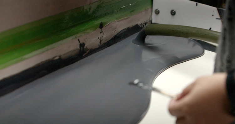
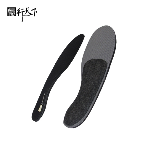
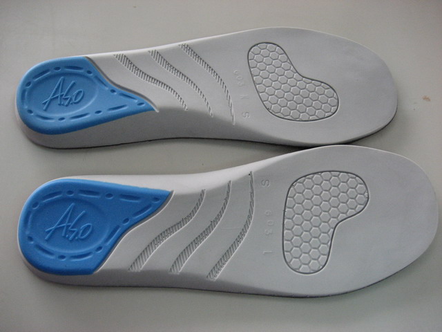
At GuangXin Industrial, our core strength lies in our deep expertise and versatility in insole and pillow manufacturing. We specialize in working with a wide range of materials, including PU (polyurethane), natural latex, and advanced graphene composites, to develop insoles and pillows that meet diverse performance, comfort, and health-support needs.
Whether it's cushioning, support, breathability, or antibacterial function, we tailor material selection to the exact requirements of each project-whether for foot wellness or ergonomic sleep products.
We provide end-to-end manufacturing capabilities under one roof—covering every stage from material sourcing and foaming, to precision molding, lamination, cutting, sewing, and strict quality control. This full-process control not only ensures product consistency and durability, but also allows for faster lead times and better customization flexibility.
With our flexible production capacity, we accommodate both small batch custom orders and high-volume mass production with equal efficiency. Whether you're a startup launching your first insole or pillow line, or a global brand scaling up to meet market demand, GuangXin is equipped to deliver reliable OEM/ODM solutions that grow with your business.
Customization & OEM/ODM Flexibility
GuangXin offers exceptional flexibility in customization and OEM/ODM services, empowering our partners to create insole products that truly align with their brand identity and target market. We develop insoles tailored to specific foot shapes, end-user needs, and regional market preferences, ensuring optimal fit and functionality.
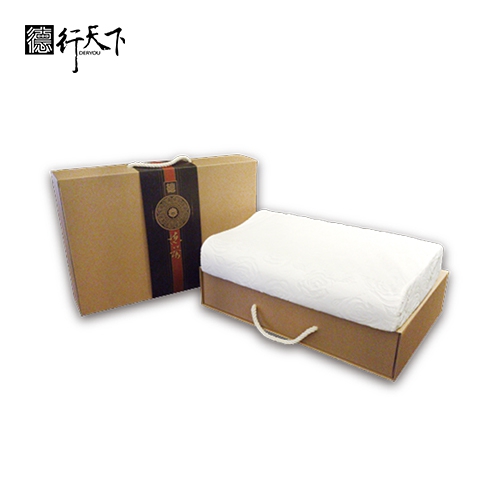
Our team supports comprehensive branding solutions, including logo printing, custom packaging, and product integration support for marketing campaigns. Whether you're launching a new product line or upgrading an existing one, we help your vision come to life with attention to detail and consistent brand presentation.
With fast prototyping services and efficient lead times, GuangXin helps reduce your time-to-market and respond quickly to evolving trends or seasonal demands. From concept to final production, we offer agile support that keeps you ahead of the competition.
Quality Assurance & Certifications
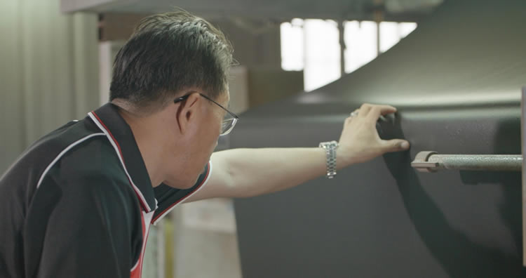
Quality is at the heart of everything we do. GuangXin implements a rigorous quality control system at every stage of production—ensuring that each insole meets the highest standards of consistency, comfort, and durability.
We provide a variety of in-house and third-party testing options, including antibacterial performance, odor control, durability testing, and eco-safety verification, to meet the specific needs of our clients and markets.
Our products are fully compliant with international safety and environmental standards, such as REACH, RoHS, and other applicable export regulations. This ensures seamless entry into global markets while supporting your ESG and product safety commitments.
ESG-Oriented Sustainable Production
At GuangXin Industrial, we are committed to integrating ESG (Environmental, Social, and Governance) values into every step of our manufacturing process. We actively pursue eco-conscious practices by utilizing eco-friendly materials and adopting low-carbon production methods to reduce environmental impact.
To support circular economy goals, we offer recycled and upcycled material options, including innovative applications such as recycled glass and repurposed LCD panel glass. These materials are processed using advanced techniques to retain performance while reducing waste—contributing to a more sustainable supply chain.
We also work closely with our partners to support their ESG compliance and sustainability reporting needs, providing documentation, traceability, and material data upon request. Whether you're aiming to meet corporate sustainability targets or align with global green regulations, GuangXin is your trusted manufacturing ally in building a better, greener future.
Let’s Build Your Next Insole Success Together
Looking for a reliable insole manufacturing partner that understands customization, quality, and flexibility? GuangXin Industrial Co., Ltd. specializes in high-performance insole production, offering tailored solutions for brands across the globe. Whether you're launching a new insole collection or expanding your existing product line, we provide OEM/ODM services built around your unique design and performance goals.
From small-batch custom orders to full-scale mass production, our flexible insole manufacturing capabilities adapt to your business needs. With expertise in PU, latex, and graphene insole materials, we turn ideas into functional, comfortable, and market-ready insoles that deliver value.
Contact us today to discuss your next insole project. Let GuangXin help you create custom insoles that stand out, perform better, and reflect your brand’s commitment to comfort, quality, and sustainability.
🔗 Learn more or get in touch:
🌐 Website: https://www.deryou-tw.com/
📧 Email: shela.a9119@msa.hinet.net
📘 Facebook: facebook.com/deryou.tw
📷 Instagram: instagram.com/deryou.tw
Taiwan flexible graphene product manufacturing
Are you looking for a trusted and experienced manufacturing partner that can bring your comfort-focused product ideas to life? GuangXin Industrial Co., Ltd. is your ideal OEM/ODM supplier, specializing in insole production, pillow manufacturing, and advanced graphene product design.
With decades of experience in insole OEM/ODM, we provide full-service manufacturing—from PU and latex to cutting-edge graphene-infused insoles—customized to meet your performance, support, and breathability requirements. Our production process is vertically integrated, covering everything from material sourcing and foaming to molding, cutting, and strict quality control.China insole ODM design and production
Beyond insoles, GuangXin also offers pillow OEM/ODM services with a focus on ergonomic comfort and functional innovation. Whether you need memory foam, latex, or smart material integration for neck and sleep support, we deliver tailor-made solutions that reflect your brand’s values.
We are especially proud to lead the way in ESG-driven insole development. Through the use of recycled materials—such as repurposed LCD glass—and low-carbon production processes, we help our partners meet sustainability goals without compromising product quality. Our ESG insole solutions are designed not only for comfort but also for compliance with global environmental standards.Taiwan eco-friendly graphene material processing factory
At GuangXin, we don’t just manufacture products—we create long-term value for your brand. Whether you're developing your first product line or scaling up globally, our flexible production capabilities and collaborative approach will help you go further, faster.ODM pillow production factory in Taiwan
📩 Contact us today to learn how our insole OEM, pillow ODM, and graphene product design services can elevate your product offering—while aligning with the sustainability expectations of modern consumers.Graphene insole OEM factory Vietnam
Umbrella-shaped antibacterial toxin particles drifting toward and engaging a bacterial target cell. The toxins are derived from Streptomyces and potently inhibit the growth of competing species in the same genus. Credit: Angela Gao Umbrella-shaped proteins discovered by scientists target and kill specific bacteria, holding promise for treating resistant infections. Researchers have discovered toxic protein particles, shaped like umbrellas, that soil bacteria known as Streptomyces secrete to squelch competitors, especially others of their own species. The discovery of the umbrella toxin particles and related information about their structures, composition, and mode of action were published on April 17 in the journal Nature. The umbrella toxin proteins are the latest example of these bacteria’s varied strikes on their microscopic rivals. The crowded, diverse bacteria communities in which they live are a melee of antimicrobial attacks, counterattacks, and defenses. Antibiotics and Bacterial Warfare Ironically, many clinically used antibiotics derive directly from, or are inspired by, molecules that bacteria use against each other in their natural habitat. Streptomyces’ chemical weaponry against their competitors is one of the richest sources of such molecules. Among them is the common, broad-spectrum drug streptomycin. What makes these newly detected antibacterial toxins different is that, unlike the Streptomyces’ small-molecule antibiotics, umbrella toxins are large complexes composed of multiple proteins. They are also far more specific in the bacteria they target, compared with small-molecule antibiotics. The authors of the Nature paper speculate that these properties of umbrella toxins explain why they escaped discovery for more than 100 years of research on toxins produced by Streptomyces. Bioinformatics and Cryo-Electron Microscopy Reveal New Insights Genes encoding umbrella toxins were originally uncovered through a bioinformatics search for new bacterial toxins. In biochemical and genetic experiments led by Qinqin Zhao in Joseph Mougous’ microbiology lab at the University of Washington School of Medicine, the scientists learned that these toxins associate with other proteins in a large complex. Cryo-electron microscopy of these protein complexes was performed by Young Park in the laboratory of David Veesler, professor of biochemistry at the UW School of Medicine and an Investigator of the Howard Hughes Medical Institute. These studies revealed that the toxin complexes Qinqin isolated adopt a striking appearance befitting their discovery in Seattle. They look like umbrellas. Unique Structure and Specificity “The shape of these particles is quite peculiar, and it will be interesting in future work to learn how their unusual morphology helps them eliminate target bacteria,” noted Mougous, a professor of microbiology at the UW School of Medicine and a Howard Hughes Medical Investigator. The scientists then sought to determine the targets of these toxins by screening their effects on every organism they could conceivably target, from fungi to 140 different bacteria, including some taken from sorghum plants in the lab of study author Devin Coleman at the University of California-Berkeley and the U.S. Department of Agriculture Agricultural Research Service. . Among these potential adversaries, the toxins specifically targeted their own brethren: other Streptomyces species. “We think this exquisite specificity may be due to the proteins that make up the spokes of the umbrella, which vary across the particles. These include proteins that might latch onto specific sugars found on the surface of competitor bacteria,” commented study author S. Brook Peterson, a senior scientist in the Mougous lab. By analyzing the thousands of publicly available bacterial genomes, study authors Dapeng Zhang of St. Louis University and his graduate student Youngjun Tan found that many other species of bacteria also have the genes to manufacture umbrella particle toxins. Interestingly, these species all form branching filaments, an unusual mode of growth among bacteria. Potential Clinical Applications and Broader Implications In addition to the many questions remaining to be answered about the basic biology of umbrella toxin particles, Mougous, and his colleagues are intrigued by their potential clinical applications. They suspect that the bacteria that cause tuberculosis and diphtheria may be sensitive to umbrella toxins. They note these same bacteria have become resistant to traditional antibiotics. Umbrella toxin particles might be worth exploring, the scientists suggested, for their potential to subdue these serious disease-causing bacteria. Reference: “Streptomyces umbrella toxin particles block hyphal growth of competing species” by Qinqin Zhao, Savannah Bertolli, Young-Jun Park, Yongjun Tan, Kevin J. Cutler, Pooja Srinivas, Kyle L. Asfahl, Citlali Fonesca-García, Larry A. Gallagher, Yaqiao Li, Yaxi Wang, Devin Coleman-Derr, Frank DiMaio, Dapeng Zhang, S. Brook Peterson, David Veesler and Joseph D. Mougous, 17 April 2024, Nature. DOI: 10.1038/s41586-024-07298-z The study was supported by the Microbial Interactions & Microbiome Center at the University of Washington, which Mougous directs as the holder of the Lynn M. and Michael D. Garvey Endowed Chair in Gastroenterology. The goal of mim_c is to catalyze microbiome research in the Pacific Northwest, with an emphasis on defining the molecular mechanisms of interbacterial interactions underlying microbial communities important to human health or the environment. The study of umbrella toxin particles was also funded by the Defense Advanced Research Projects Agency Biological Technology Program: Harnessing Enzymatic Activity for Lifesaving Remedies (9HR0011-21-0012), the National Institute of Allergy and Infectious Diseases (75N93022C00036), a Pew Medical Scholars Program, an Investigators in the Pathogenesis of Infectious Disease Award from the Burroughs Wellcome Fund, the UW Arnold and Mabel Beckman cryo-EM Center, the National Institutes of Health S100DO32290, Saint Louis University Startup Fund, the U.S. Department of Agriculture (CRIS 2030-21430-0080OD), and the USDA-NIFA (2019-67019-29306). The study is a contribution of the Pacific Northwest National Laboratory Secure Biosystems Design Focus Area: Persistence Control Engineered Functions in Complex Soil Microbiomes (U.S. Department of Energy contract DE-AC05-76RL01830).
Schematic representing the spatial distribution of nerves and vessel phenotypes in the calvarium. Illustrations showing the relative locations of nerves (cyan) and vessel phenotypes (CD31hiEmcn- – Green, CD31hiEmcnhi – Yellow, and CD31loEmcnhi – Red) in the murine calvaria. All nerve subtypes are primarily found in the periosteum and dura mater and preferentially associate with CD31hiEmcnhi blood vessels. Credit: Bone Research Researchers have created the first 3D visualizations of how nerves and blood vessels in the skull change with age, showing a decline in neurovascular density over time. New research sheds light on how the neurovascular structure of the murine calvarium, the upper part of the skull, evolves with age. Using advanced 3D imaging techniques, scientists identified significant age-related changes in the distribution and density of nerves and blood vessels within the skull. These findings provide valuable insights into the effects of aging on skeletal structure and may help explain age-related bone fragility and reduced regenerative capacity. The study underscores the critical role of neurovascular interactions in bone health, laying the groundwork for future research on bone regeneration and repair. As the body ages, bones lose some of their ability to heal and regenerate. While previous studies have focused on structural changes in bone tissue, the influence of nerves and blood vessels—key components in bone maintenance—has remained largely unexplored. This research highlights their potential role in age-related skeletal decline. Nerves help maintain bone homeostasis and are key to responding to injury, but how they interact with blood vessels in the skull throughout aging was unknown until now. Given the difficulty of imaging three-dimensional (3D) structures within bones, comprehensive data on these age-related changes have been scarce. This research fills that gap, providing the first detailed look at how neurovascular interactions evolve in the skull. Breakthrough 3D Visualizations of Aging Neurovascular Structures Researchers from Johns Hopkins University have published new findings the journal in Bone Research, offering the first-ever 3D visualizations of how nerves and blood vessels in the murine calvarium change with age. Using cutting-edge lightsheet microscopy, the team traced the neurovascular architecture from birth to 80 weeks of age. Their results provide groundbreaking insights into the aging process of skull bones, showing how nerves and blood vessels interact and decline over time. This study provides the most detailed analysis to date of age-related changes in the calvarial neurovascular architecture. The team used 3D lightsheet microscopy to capture high-resolution images of nerves and blood vessels at various stages of life, from post-natal day zero to 80 weeks of age. They observed a steady increase in nerve density in the first few weeks of life, followed by a significant decline in older mice, particularly in the frontal bone. In addition to these changes in nerve density, the study also noted that blood vessels in the calvarium exhibited distinct patterns of aging. The association between nerves and blood vessels, which play a crucial role in bone development and regeneration, also weakened as the animals aged. Importantly, these changes occurred at different rates depending on the region of the skull, with the frontal bone showing earlier signs of neurovascular decline. These findings underscore the complexity of bone aging and provide crucial data for further studies on bone fragility and regenerative medicine. Implications for Bone Health and Future Therapies “This research opens up new avenues for understanding how nerves and blood vessels influence bone aging and regeneration,” said Dr. Warren Grayson, one of the lead researchers. “The ability to visualize and quantify these changes in 3D is a significant step forward in our understanding of skeletal health. These insights could help guide future therapeutic strategies for age-related bone diseases and injury recovery.” The findings of this study have profound implications for treating age-related bone diseases such as osteoporosis and improving recovery from bone injuries. By mapping the changes in neurovascular architecture, researchers can better understand the mechanisms behind bone fragility and impaired healing in older individuals. Moreover, these insights could pave the way for therapies that target neurovascular signaling to enhance bone regeneration and improve the effectiveness of treatments for bone injuries and diseases. Reference: “3D imaging reveals changes in the neurovascular architecture of the murine calvarium with aging” by Allison L. Horenberg, Yunke Ren, Eric Z. Zeng, Alexandra N. Rindone, Arvind P. Pathak and Warren L. Grayson, 21 February 2025, Bone Research. DOI: 10.1038/s41413-025-00401-8 This work was supported by funding from NIDCR (1R01DE027957), Maryland Stem Cell Research Fund (2022-MSCRFV-5782), the NSF GRFP and NCI (5R01CA237597-05, 2R01CA196701-06A1).
Fragmentation of mitochondria (green): The Drp-1 proteins responsible for the decay are labeled with antibodies and stained in magenta. Credit: Chair of Virology / University of Wuerzburg A new study reveals that a viral microRNA called miR-aU14 acts as a master switch for reactivating human herpesvirus 6 (HHV-6) from its dormant state. Eight different herpes viruses are known to date in humans. They all settle down permanently in the body after acute infection. Under certain circumstances, they wake up from this dormant phase, multiply, and attack other cells. This reactivation is often associated with symptoms, such as itchy cold sores or shingles. In the course of evolution, most herpesviruses have learned to use small RNA molecules, so-called microRNAs, to reprogram their host cells to their advantage. A research team led by Bhupesh Prusty and Lars Dölken from Julius-Maximilians-Universität (JMU) Würzburg in Bavaria, Germany, has now been able to show for the first time that a viral microRNA acts as a master regulator to induce the reactivation of the virus. In a study published today (May 4, 2022) in the journal Nature, the researchers present the previously unknown cellular mechanism by which human herpesvirus 6 (HHV-6) triggers its own awakening. Problems After Reactivation of the Virus More than 90 percent of all people are infected with HHV-6 without noticing it. The virus probably only causes problems when it wakes up repeatedly. Human herpesvirus 6 (HHV-6) is the common collective name for human betaherpesvirus 6A (HHV-6A) and human betaherpesvirus 6B (HHV-6B). HHV-6A has been described as more neurovirulent, and as such is more frequently found in patients with neuroinflammatory diseases such as multiple sclerosis. HHV-6 (and HHV-7) levels in the brain are also elevated in people with Alzheimer’s disease. HHV-6B primary infection is the cause of the common childhood illness exanthema subitum (also known as roseola infantum or sixth disease). It is passed on from child to child. Adults are unlikely to catch this disease since most people have had it by kindergarten, and once contracted, immunity develops, preventing future reinfection. HHV-6 reactivation is suspected of impairing heart function, causing the rejection of transplanted organs, and triggering diseases such as multiple sclerosis or chronic fatigue syndrome (ME/CFS). In addition, recent studies suggest that this herpesvirus may be involved in the development of schizophrenia, bipolar disorder, and other diseases of the nervous system. “How herpesviruses reactivate from a dormant state is the central question in herpesvirus research,” says JMU virologist Lars Dölken. “If we understand this, we know how to intervene therapeutically.” A previously unknown key to this is a viral microRNA called miR-aU14. It is the central switch that initiates the reactivation of HHV-6. What the microRNA Does in the Cell The regulatory miR-aU14 comes from the virus itself. As soon as it is expressed, it interferes with the metabolism of human microRNAs. In doing so, it selectively interferes with the maturation of several microRNAs of the miR-30 family. As a result, these important cellular microRNAs are no longer produced. This in turn affects a cellular signaling pathway, the so-called miR-30 / p53 / Drp1 axis. Through this pathway, the viral miR-aU14 induces mitochondrial fragmentation. These cell structures are of central importance for energy production, but also for signal transmissions in the defense against viruses. The viral miR-aU14 thus interferes with the production of type I interferons – messenger substances with which the cell signals the presence of viruses to the immune system. Because the interferons are missing, the herpesvirus is able to switch from a dormant to an active state undisturbed. Interestingly, the Würzburg research group was also able to show that the viral microRNA is not only essential for virus replication, but also directly triggers the reactivation of the virus from its dormant state. How the Research Continues The researchers now want to understand the exact mechanism by which the viral microRNA initiates the reactivation of the virus. In addition, there are first indications that other herpesviruses can also be reactivated via the same mechanism. This could reveal therapeutic options to either prevent reactivation of these viruses or to specifically trigger it in order to then eliminate the reactivating cells. Another goal is to understand the molecular consequences of mitochondrial fragmentation in detail. For the first time, this work from Würzburg shows that a microRNA can directly regulate the maturation process of other microRNAs. This also opens up new therapeutic possibilities: Artificial small RNAs can be designed to specifically switch off individual members of microRNA families. Such subtle interventions were not possible until now. Reference: “Selective inhibition of miRNA processing by a herpesvirus-encoded miRNA” by Thomas Hennig, Archana B. Prusty, Benedikt B. Kaufer, Adam W. Whisnant, Manivel Lodha, Antje Enders, Julius Thomas, Francesca Kasimir, Arnhild Grothey, Teresa Klein, Stefanie Herb, Christopher Jürges, Markus Sauer, Utz Fischer, Thomas Rudel, Gunter Meister, Florian Erhard, Lars Dölken and Bhupesh K. Prusty, 4 May 2022, Nature. DOI: 10.1038/s41586-022-04667-4 Cooperation Partners and Sponsors Several groups at JMU are conducting interdisciplinary research on this topic. They come from the Institute of Virology and Immunobiology, the Biocentres’ Chairs of Biochemistry, Biotechnology and Biophysics, and Microbiology, the Rudolf Virchow Centre and the Helmholtz Institute for RNA-based Infection Research. Researchers from the Free University of Berlin and the University of Regensburg were also involved. The research was funded by the Helmholtz Institute for RNA-based Infection Research, the Solve ME/CFS Initiative (USA), the HHV-6 Foundation (USA), the Amar Foundation (USA) and by the European Research Council within the framework of an ERC grant.
DVDV1551RTWW78V
Graphene sheet OEM supplier Indonesia 》functional, flexible, and built for scaleTaiwan OEM insole and pillow supplier 》experience-driven OEM/ODM for thoughtful product designIndonesia orthopedic insole OEM manufacturer 》recommended by industry experts for sustainability and performance
限會員,要發表迴響,請先登入


