Introduction – Company Background
GuangXin Industrial Co., Ltd. is a specialized manufacturer dedicated to the development and production of high-quality insoles.
With a strong foundation in material science and footwear ergonomics, we serve as a trusted partner for global brands seeking reliable insole solutions that combine comfort, functionality, and design.
With years of experience in insole production and OEM/ODM services, GuangXin has successfully supported a wide range of clients across various industries—including sportswear, health & wellness, orthopedic care, and daily footwear.
From initial prototyping to mass production, we provide comprehensive support tailored to each client’s market and application needs.
At GuangXin, we are committed to quality, innovation, and sustainable development. Every insole we produce reflects our dedication to precision craftsmanship, forward-thinking design, and ESG-driven practices.
By integrating eco-friendly materials, clean production processes, and responsible sourcing, we help our partners meet both market demand and environmental goals.
Core Strengths in Insole Manufacturing
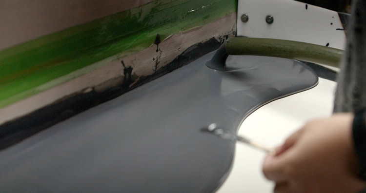
At GuangXin Industrial, our core strength lies in our deep expertise and versatility in insole and pillow manufacturing. We specialize in working with a wide range of materials, including PU (polyurethane), natural latex, and advanced graphene composites, to develop insoles and pillows that meet diverse performance, comfort, and health-support needs.
Whether it's cushioning, support, breathability, or antibacterial function, we tailor material selection to the exact requirements of each project-whether for foot wellness or ergonomic sleep products.
We provide end-to-end manufacturing capabilities under one roof—covering every stage from material sourcing and foaming, to precision molding, lamination, cutting, sewing, and strict quality control. This full-process control not only ensures product consistency and durability, but also allows for faster lead times and better customization flexibility.
With our flexible production capacity, we accommodate both small batch custom orders and high-volume mass production with equal efficiency. Whether you're a startup launching your first insole or pillow line, or a global brand scaling up to meet market demand, GuangXin is equipped to deliver reliable OEM/ODM solutions that grow with your business.
Customization & OEM/ODM Flexibility
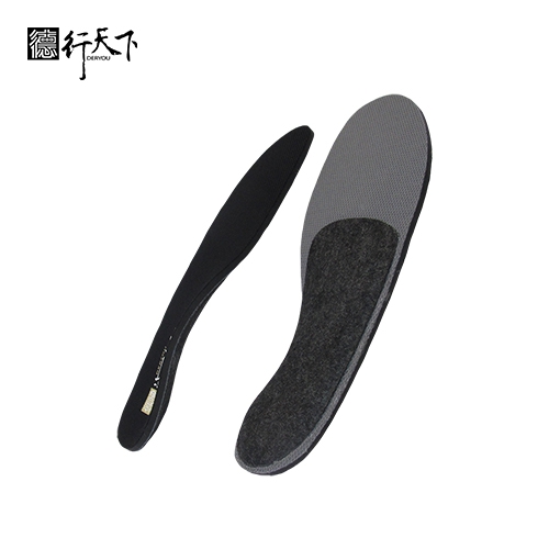
GuangXin offers exceptional flexibility in customization and OEM/ODM services, empowering our partners to create insole products that truly align with their brand identity and target market. We develop insoles tailored to specific foot shapes, end-user needs, and regional market preferences, ensuring optimal fit and functionality.
Our team supports comprehensive branding solutions, including logo printing, custom packaging, and product integration support for marketing campaigns. Whether you're launching a new product line or upgrading an existing one, we help your vision come to life with attention to detail and consistent brand presentation.
With fast prototyping services and efficient lead times, GuangXin helps reduce your time-to-market and respond quickly to evolving trends or seasonal demands. From concept to final production, we offer agile support that keeps you ahead of the competition.
Quality Assurance & Certifications
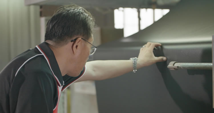
Quality is at the heart of everything we do. GuangXin implements a rigorous quality control system at every stage of production—ensuring that each insole meets the highest standards of consistency, comfort, and durability.
We provide a variety of in-house and third-party testing options, including antibacterial performance, odor control, durability testing, and eco-safety verification, to meet the specific needs of our clients and markets.
Our products are fully compliant with international safety and environmental standards, such as REACH, RoHS, and other applicable export regulations. This ensures seamless entry into global markets while supporting your ESG and product safety commitments.
ESG-Oriented Sustainable Production
At GuangXin Industrial, we are committed to integrating ESG (Environmental, Social, and Governance) values into every step of our manufacturing process. We actively pursue eco-conscious practices by utilizing eco-friendly materials and adopting low-carbon production methods to reduce environmental impact.
To support circular economy goals, we offer recycled and upcycled material options, including innovative applications such as recycled glass and repurposed LCD panel glass. These materials are processed using advanced techniques to retain performance while reducing waste—contributing to a more sustainable supply chain.
We also work closely with our partners to support their ESG compliance and sustainability reporting needs, providing documentation, traceability, and material data upon request. Whether you're aiming to meet corporate sustainability targets or align with global green regulations, GuangXin is your trusted manufacturing ally in building a better, greener future.
Let’s Build Your Next Insole Success Together
Looking for a reliable insole manufacturing partner that understands customization, quality, and flexibility? GuangXin Industrial Co., Ltd. specializes in high-performance insole production, offering tailored solutions for brands across the globe. Whether you're launching a new insole collection or expanding your existing product line, we provide OEM/ODM services built around your unique design and performance goals.
From small-batch custom orders to full-scale mass production, our flexible insole manufacturing capabilities adapt to your business needs. With expertise in PU, latex, and graphene insole materials, we turn ideas into functional, comfortable, and market-ready insoles that deliver value.
Contact us today to discuss your next insole project. Let GuangXin help you create custom insoles that stand out, perform better, and reflect your brand’s commitment to comfort, quality, and sustainability.
🔗 Learn more or get in touch:
🌐 Website: https://www.deryou-tw.com/
📧 Email: shela.a9119@msa.hinet.net
📘 Facebook: facebook.com/deryou.tw
📷 Instagram: instagram.com/deryou.tw
Innovative pillow ODM solution in China
Are you looking for a trusted and experienced manufacturing partner that can bring your comfort-focused product ideas to life? GuangXin Industrial Co., Ltd. is your ideal OEM/ODM supplier, specializing in insole production, pillow manufacturing, and advanced graphene product design.
With decades of experience in insole OEM/ODM, we provide full-service manufacturing—from PU and latex to cutting-edge graphene-infused insoles—customized to meet your performance, support, and breathability requirements. Our production process is vertically integrated, covering everything from material sourcing and foaming to molding, cutting, and strict quality control.Innovative pillow ODM solution in Indonesia
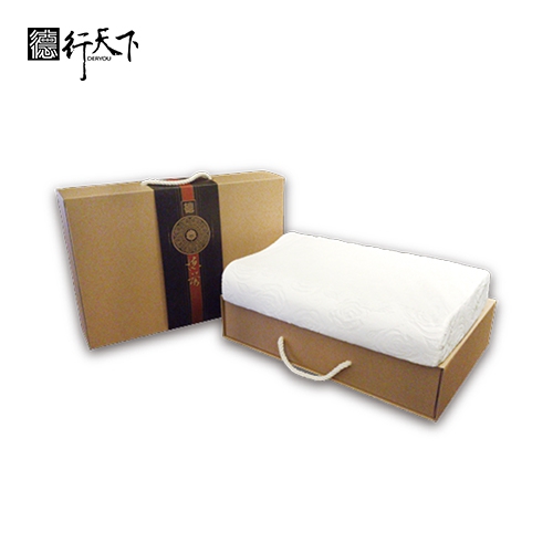
Beyond insoles, GuangXin also offers pillow OEM/ODM services with a focus on ergonomic comfort and functional innovation. Whether you need memory foam, latex, or smart material integration for neck and sleep support, we deliver tailor-made solutions that reflect your brand’s values.
We are especially proud to lead the way in ESG-driven insole development. Through the use of recycled materials—such as repurposed LCD glass—and low-carbon production processes, we help our partners meet sustainability goals without compromising product quality. Our ESG insole solutions are designed not only for comfort but also for compliance with global environmental standards.Cushion insole OEM solution Vietnam
At GuangXin, we don’t just manufacture products—we create long-term value for your brand. Whether you're developing your first product line or scaling up globally, our flexible production capabilities and collaborative approach will help you go further, faster.Indonesia graphene product OEM service
📩 Contact us today to learn how our insole OEM, pillow ODM, and graphene product design services can elevate your product offering—while aligning with the sustainability expectations of modern consumers.Orthopedic pillow OEM solutions Vietnam
Researchers have uncovered an evolutionary “tipping point” affecting fungi growth and shape diversity, highlighting how minor environmental changes can significantly impact evolution. This discovery, based on studies of fungi and water molds, reveals that certain hyphal shapes are favored due to evolutionary constraints. The findings offer new insights into the resilience of species to climate change and the potential for creating targeted antimicrobials against fungal pathogens. Research shows small environmental changes can significantly influence the structures of cells and organisms. Scientists have found a “tipping point” in the evolution of fungi that throttles their growth and sculpts their shapes. The findings, published in the journal Cell Reports, demonstrate how small changes in environmental factors can lead to huge changes in evolutionary outcomes. Fungi are nature’s great composters. They wait within the forest floor to feed on fallen trees and autumn leaves, releasing essential nutrients from these plants back into the Earth. Hyphae of the fungus Allomyces arbuscula growing as part of a mycelium. Credit: Maxim Ohairwe Although fungi often bring to mind mushroom caps, fungi also have underground “roots” called mycelia. Mycelia are made up of thousands of interconnected, microscopic, finger-like cells called hyphae that grow into vast networks. Hyphae worm their way through the soil by growing from their tips. To do so, they inflate themselves, similar to the long balloons used to make balloon animals. Their elongated forms allow hyphae to locate and consume nutrients within the soil. But not all hyphae are the same shape: some have rounded tips, while others are pointed. The hyphae of water molds—fungus-like pathogens that cause blight in crops—are particularly pointy. The fitness landscape of hyphae showing that natural shapes are constrained by a tipping point. Credit: Maxim Ohairwe Exploring Hyphal Shapes “A major challenge in biology is to identify the specific evolutionary factors that determine the shape—or form—of a given organism,” said Enrique Rojas, assistant professor of biology at New York University and the study’s senior author. To understand the reasons for different shapes of hyphae, Rojas and his colleagues combined theory and experiments to investigate fungi and water molds from across nature. They first employed physics-based models of inflationary tip growth to determine all possible shapes of hyphae. Surprisingly, the shapes of actual hyphae found in nature assumed only a small subset of the possible shapes. An Achlya bisexualis hypha growing. Credit: Maxim Ohairwe The researchers hypothesized that the limited shapes observed in nature reflected “survival of the fittest,” and that the many possible shapes not observed in real fungi were, for some reason, weaker evolutionary rejects. To explore this idea, they examined the growth rate of hyphae with different shapes to create a fitness landscape for hyphae. “Our eureka moment was when we realized that the shapes of hyphae were intimately connected to their ability to grow fast,” said Maxim Ohairwe, a PhD student in NYU’s Department of Biology and the lead author of the study. Hyphal mycelium. Credit: Maxim Ohairwe Fitness Landscapes and Evolution A fitness landscape is like a topographic map that visualizes the evolution of an organism: every species wanders through its fitness landscape by testing whether or not random mutations in its genes increase its growth rate, or fitness. A species only stops its restless wandering when a new mutation decreases its fitness—that is, when it is at a fitness peak. However, Rojas’s team found that fitness landscapes can be much more rich than a system of peaks and valleys. In fact, they found that the fitness landscape for hyphae contained an overhanging cliff, or tipping point, and that this acts as a barrier to evolution, strongly limiting the shapes of fungal hyphae. Accordingly, they predicted that hyphae with shapes near the brink of the tipping point would be particularly vulnerable to small environmental, chemical, or genetic changes. An Achlya bisexualis hypha that was treated with a chemical that blocks subcellular transport. The treatment caused the hypha to elongate much more slowly and with a strange nub shape not found in nature. Credit: Maxim Ohairwe The researchers tested their prediction by treating fungi near the tipping point with small amounts of chemicals that affected hyphal growth. They used one chemical that reduces pressure within the hyphae and another derived from a sea sponge that blocks the hypha’s ability to deliver cellular components to the tip of the cell. Both treatments caused the same dramatic effect: the hyphae elongated much more slowly and with a strange nub shape not found in nature. “Our findings explain hyphal shape diversity in an enormous, diverse, and important group of species,” said Rojas. “More broadly, they also demonstrate an important new evolutionary principle: that fitness landscapes can have instabilities, or tipping points, that impose strict constraints on complex traits, like biological form.” The researchers believe that their results have critical implications for our understanding of many ecological and evolutionary systems. For example, those species whose evolution is subject to a tipping point may be the most vulnerable to the gradual increase in temperature caused by climate change. Their findings could also aid in the development of new antimicrobials against disease-causing fungi by identifying vulnerabilities in their growth associated with an evolutionary tipping point. Reference: “A fitness landscape instability governs the morphological diversity of tip-growing cells” by Maxim E. Ohairwe, Branka D. Živanović and Enrique R. Rojas, 25 March 2024, Cell Reports. DOI: 10.1016/j.celrep.2024.113961 In addition to Rojas and Ohairwe, Branka Živanović of the University of Belgrade was a study coauthor. This research was supported by the National Science Foundation (CAREER grant 2047404, grant PHY-221045), the Ministry of Education, Science and Technological Development of the Republic of Serbia (451-03-68/2022-14/200053), and Deutsche Forschungsgemeinschaft (GA 173/13-1). The NYU Microscopy Laboratory, where electron microscopy was performed, is funded by the National Institutes of Health (NCI P30CA016087 and S10 OD019974).
First author and scientific diver Erin Shilling treats a diseased stony coral affected by stony coral tissue loss disease. Credit: Joshua Voss, Ph.D., FAU Harbor Branch, Coral Reef and Health Ecology Lab FAU Harbor Branch Oceanographic Institute study shows 95 percent success rate with amoxicillin. Diseases continue to be a major threat to coral reef health. For example, a relatively recent outbreak termed stony coral tissue loss disease is an apparently infectious waterborne disease known to affect at least 20 stony coral species. First discovered in 2014 in Miami-Dade County, the disease has since spread throughout the majority of Florida’s Coral Reef and into multiple countries and territories in the Caribbean. Some reefs of the northern section of Florida’s Coral Reef are experiencing as much as a 60 percent loss of living coral tissue area. A new study by researchers at Florida Atlantic University’s Harbor Branch Oceanographic Institute reveals how a common antibiotic used to treat bacterial infections in humans is showing promise in treating disease-affected Montastraea cavernosa coral colonies in situ. M. cavernosa, also known as the Great Star Coral, is a hard or stony coral found widely throughout the tropical western Atlantic, including several regions currently affected by stony coral tissue loss disease. Preserving M. cavernosa colonies is of particular importance due to its high abundance and role as a dominant reef builder in the northern section of Florida’s Coral Reef. FAU scientific divers Erin Shilling and Ryan Eckert are shown applying the antibiotic treatment (the white paste) into trenches created around disease lesions present at the edges of the coral colony. The trenches are the white inner margin on the coral tissue (the white is just exposed coral skeleton), while the lesions are the pale/white tissue at the edge of the coral colony. Shilling and Eckert then apply the same treatment to a smaller coral in the same manner. Later, they again treat a coral, however, it is with chlorinated epoxy (brown paste) being packed into both the trench created on the colony as well as over the lesions themselves. Credit: Joshua Voss, Ph.D., FAU Harbor Branch, Coral Reef and Health Ecology Lab The objective of the study, published in Scientific Reports, was to experimentally assess the effectiveness of two intervention treatments: chlorinated epoxy and amoxicillin combined with Core Rx/Ocean Alchemists Base 2B as compared to untreated controls. Results showed that the Base 2B plus amoxicillin treatment had a 95 percent success rate at healing individual disease lesions. However, it did not necessarily prevent treated colonies from developing new lesions over time. Chlorinated epoxy treatments were not significantly different from untreated control colonies, suggesting that chlorinated epoxy treatments are an ineffective intervention technique for stony coral tissue loss disease. “There are three possible scenarios that may explain the appearance of new lesions in the amoxicillin treated lesions of the corals that had healed in our study,” said Erin N. Shilling, M.S., first author and a recent graduate of the Marine Science and Oceanography master degree program at FAU Harbor Branch. “It’s possible that the causative agent of stony coral tissue loss disease is still present in the environment and is re-infecting quiesced colonies. It also could be that the duration and dose of this antibiotic intervention was sufficient to arrest stony coral tissue loss disease at treated lesions, but insufficient at eliminating its pathogens from other areas of the coral colony.” A stony coral treated with the Base 2B plus amoxicillin treatment. Credit: Joshua Voss, Ph.D., FAU Harbor Branch, Coral Reef and Health Ecology Lab The study was conducted approximately 2 kilometers offshore from Lauderdale-by-the-Sea in Broward County, Florida, at sites with a maximum depth of 10 meters. Both colony disease status and treated lesion status were analyzed independently so that the treatment’s effectiveness at halting individual lesions could be assessed while also determining if a treatment had any impact on the colony as a whole. Colonies were monitored periodically over 11 months to assess treatment effectiveness by tracking lesion development and overall disease status. “Success in treating stony coral tissue loss disease with antibiotics may benefit from using approaches typically successful against bacterial infections in humans, for example using a strong initial dose of antibiotics followed by a regimen of smaller supplementary doses over time,” said Joshua Voss, Ph.D., senior author, an associate research professor at FAU Harbor Branch and executive director of the NOAA Cooperative Institute for Ocean Exploration, Research, and Technology. “Future research efforts should focus on assessing the potential unintended consequences of antibiotic treatments on corals, their microbial communities, and neighboring organisms. In addition, further efforts are needed to optimize dosing and delivery methods for antibiotic treatments on stony coral tissue loss disease-affected corals and scale up intervention treatments effectively.” Voss notes that many coral diseases are still poorly characterized, which has led to calls for increased research and intervention efforts to support adaptive management strategies particularly given the considerable impacts of diseases on coral reefs over the past five decades. “Results of our experiment expand management options during coral disease outbreaks and contribute to overall knowledge regarding coral health and disease,” said Voss. This research is part of a highly coordinated collaboration through the Disease Advisory Committee (DAC) organized by the Florida Department of Environmental Protection (floridadep.gov/rcp/coral/content/stony-coral-tissue-loss-disease-response) and NOAA. Voss and Shilling are members of the DAC and part of the reconnaissance and intervention team that has collaboratively developed treatment methods, research objectives, and responses to this disease outbreak. Researchers from Nova Southeastern University, Smithsonian Marine Station and the Florida Fish and Wildlife Conservation Commission also are major partners on this team. Ian Combs, M.S., another recent FAU graduate from Voss’s lab and a co-author, helped to develop some of the coral fate-tracking techniques used in the study. “We recommend that coral reef managers and intervention specialists, particularly those focusing on stony coral tissue loss disease, adopt 3D photogrammetric methods to ensure that data are more accurate than 2D and in-water estimates,” said Combs. Reference: “Assessing the effectiveness of two intervention methods for stony coral tissue loss disease on Montastraea cavernosa” by Erin N. Shilling, Ian R. Combs and Joshua D. Voss, 21 April 2021, Scientific Reports. DOI: 10.1038/s41598-021-86926-4 This research was funded by the Florida Department of Environmental Protection (Awards B430E1 and B55008 awarded to Voss), and the Environmental Protection Agency (South Florida Geographic Initiative award X7 00D667-17). Additional funding was awarded to Shilling by the Harbor Branch Oceanographic Institute Foundation through the Indian River Lagoon Graduate Research Fellowship. All work was carried out under permission of the Florida Fish and Wildlife Conservation Commission (permits SAL-18-2022-SRP and ASL-19-1702-SRP).
Muscle stem cells and fibers can be grown in the laboratory from reprogrammed connective tissue cells (microscopy image). Credit: ETH Zurich / Bar-Nur lab ETH Zurich researchers have pioneered an mRNA-based technique for generating muscle stem cells without genetic modifications, offering new possibilities for treating muscle diseases and advancing cultured meat production. Professor Ori Bar-Nur and his colleagues at ETH Zurich are pioneering the cultivation of muscle cells in the lab, currently using mouse cells as their primary model. While their current studies are centered on mouse cells, the team is also keen on exploring the potential of human and cow cells. The implications of their research are manifold: lab-cultured human muscle tissue could serve surgical needs, while human muscle stem cells might offer therapeutic solutions for those with muscle diseases. On the other hand, cultivating cow muscle tissue in labs could transform the meat industry by eliminating the necessity of animal slaughter. For now, however, the ETH team’s research is focused on optimizing the generation of muscle stem cells and making it safer. They have now succeeded in doing so via a new approach. Reprogrammed Cells Like other researchers in the field, the scientists at ETH Zurich use a different, easier-to-grow cell type as the starting material for generating muscle cells: connective tissue cells. Using a cocktail of small molecules and proteins, they molecularly “reprogram” these cells, thereby converting them into muscle stem cells, which then multiply rapidly and produce muscle fibers. “This approach enabled us to produce large quantities of muscle cells,” explains Xhem Qabrati, a doctoral student in the Bar-Nur group and one of two lead co-authors on this study. “Although muscle cells could also be cultured directly from muscle biopsies, the cells tend to lose their functionality after isolation, rendering it challenging to produce large quantities of cells.” An important component of the used cocktail – and a central catalyst for cell transformation – is the protein MyoD. This is a transcription factor that regulates the activity of certain muscle genes in the cell nucleus. MyoD is not normally present in connective tissue cells. Before these cells can turn into muscle cells, scientists have to coax them to produce MyoD in their nucleus for several days. No Genetic Engineering Until now, researchers have turned to genetic engineering for this process: They used viral particles to carry the DNA blueprint for the MyoD protein into the cell nucleus. There, the viruses insert these building instructions into the genome, enabling the cells to produce the MyoD protein. However, this approach carries a safety risk: scientists cannot control where exactly in the genome viruses insert these instructions. Sometimes the viruses integrate into the middle of a vital gene, damaging it, or this insertion process might lead to changes that can trigger cancer cell formation. This time, Bar-Nur and his colleagues used a different approach to deliver MyoD to connective tissue cells, inspired by the mRNA vaccines for COVID-19: instead of using viruses to introduce the DNA blueprint of the MyoD gene, they introduce the mRNA transcript of this gene into cells. Since this leaves the cells’ genome unchanged, it avoids the negative consequences associated with such changes. The mRNA still enables the connective tissue cells to produce the MyoD protein, such that – together with the other components of the cocktail optimised by the ETH researchers – they can turn into muscle stem cells and fibers. The researchers recently published their new approach in the journal NPJ Regenerative Medicine. They are the first to reprogram connective tissue cells into muscle stem cells without genetic engineering. Help With Muscle Dystrophy The muscle cells produced this way are also fully functional, as the researchers have shown in experiments with mice suffering from Duchenne muscular dystrophy. In humans, this rare hereditary disease leaves sufferers lacking a protein necessary for muscle stability, meaning they experience progressive muscle wasting and paralysis. The ETH Zurich scientists injected non-defective muscle stem cells into the muscles of Duchenne muscular dystrophy mice carrying this defect. They were able to show that the healthy stem cells form repaired muscle fibers in the muscles. “Muscle stem cell transplantation of this sort could be especially helpful for patients with advanced Duchenne, who are already severely affected by muscle atrophy,” explains Inseon Kim, another doctoral student in the Bar-Nur group and a lead co-author on this study. The method is suitable for producing large quantities of muscle stem cells required for this purpose. What’s more, the fact that it does so without genetic engineering and the associated risks makes it attractive for potential future therapeutic use in humans. Alternative Meat Production However, the researchers have yet to adapt their approach to human cells; this is their next step. “In addition, we wish to investigate whether it’s also possible to convert connective tissue cells into muscle cells directly in the body by injecting the MyoD mRNA and the other cocktail components into mice affected by muscle diseases,” Bar-Nur says. This approach, too, might one day help human patients. Finally, Bar-Nur and his team would like to incorporate their new findings into their ongoing work with cow cells – another research stream of the lab. They hope this method would assist current efforts to culture animal muscle stem cells for cultivated meat production, an alternative method to produce meat for consumption. Reference: “Transgene-free direct conversion of murine fibroblasts into functional muscle stem cells” by Xhem Qabrati, Inseon Kim, Adhideb Ghosh, Nicola Bundschuh, Falko Noé, Andrew S. Palmer and Ori Bar-Nur, 8 August 2023, npj Regenerative Medicine. DOI: 10.1038/s41536-023-00317-z
DVDV1551RTWW78V
Custom graphene foam processing Vietnam 》driving your product success through every stage of manufacturingIndonesia anti-bacterial pillow ODM design 》delivering consistent quality from sample to mass productionVietnam ergonomic pillow OEM supplier 》the smart choice for brands seeking quality and customization
下一則: Custom foam pillow OEM in China 》your competitive
限會員,要發表迴響,請先登入


