Introduction – Company Background
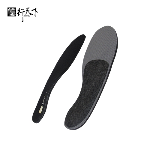
GuangXin Industrial Co., Ltd. is a specialized manufacturer dedicated to the development and production of high-quality insoles.
With a strong foundation in material science and footwear ergonomics, we serve as a trusted partner for global brands seeking reliable insole solutions that combine comfort, functionality, and design.
With years of experience in insole production and OEM/ODM services, GuangXin has successfully supported a wide range of clients across various industries—including sportswear, health & wellness, orthopedic care, and daily footwear.
From initial prototyping to mass production, we provide comprehensive support tailored to each client’s market and application needs.
At GuangXin, we are committed to quality, innovation, and sustainable development. Every insole we produce reflects our dedication to precision craftsmanship, forward-thinking design, and ESG-driven practices.
By integrating eco-friendly materials, clean production processes, and responsible sourcing, we help our partners meet both market demand and environmental goals.
Core Strengths in Insole Manufacturing
At GuangXin Industrial, our core strength lies in our deep expertise and versatility in insole and pillow manufacturing. We specialize in working with a wide range of materials, including PU (polyurethane), natural latex, and advanced graphene composites, to develop insoles and pillows that meet diverse performance, comfort, and health-support needs.
Whether it's cushioning, support, breathability, or antibacterial function, we tailor material selection to the exact requirements of each project-whether for foot wellness or ergonomic sleep products.
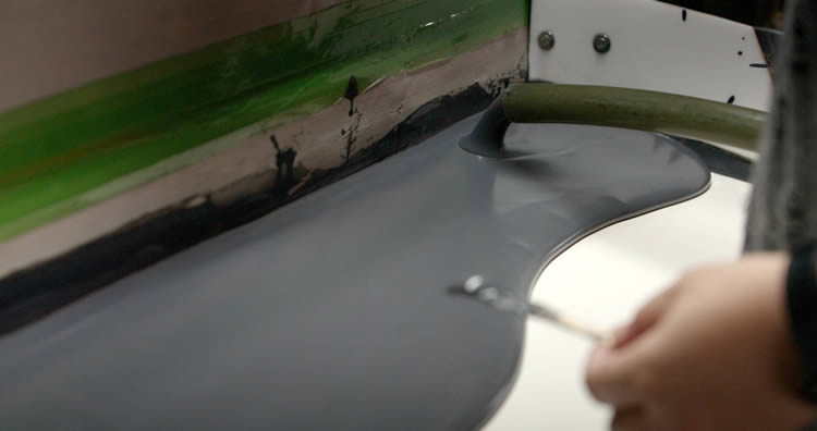
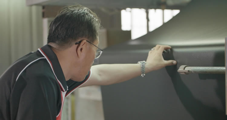
We provide end-to-end manufacturing capabilities under one roof—covering every stage from material sourcing and foaming, to precision molding, lamination, cutting, sewing, and strict quality control. This full-process control not only ensures product consistency and durability, but also allows for faster lead times and better customization flexibility.
With our flexible production capacity, we accommodate both small batch custom orders and high-volume mass production with equal efficiency. Whether you're a startup launching your first insole or pillow line, or a global brand scaling up to meet market demand, GuangXin is equipped to deliver reliable OEM/ODM solutions that grow with your business.
Customization & OEM/ODM Flexibility
GuangXin offers exceptional flexibility in customization and OEM/ODM services, empowering our partners to create insole products that truly align with their brand identity and target market. We develop insoles tailored to specific foot shapes, end-user needs, and regional market preferences, ensuring optimal fit and functionality.
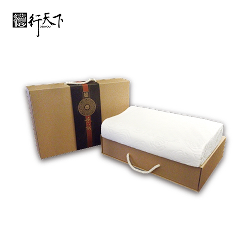
Our team supports comprehensive branding solutions, including logo printing, custom packaging, and product integration support for marketing campaigns. Whether you're launching a new product line or upgrading an existing one, we help your vision come to life with attention to detail and consistent brand presentation.
With fast prototyping services and efficient lead times, GuangXin helps reduce your time-to-market and respond quickly to evolving trends or seasonal demands. From concept to final production, we offer agile support that keeps you ahead of the competition.
Quality Assurance & Certifications
Quality is at the heart of everything we do. GuangXin implements a rigorous quality control system at every stage of production—ensuring that each insole meets the highest standards of consistency, comfort, and durability.
We provide a variety of in-house and third-party testing options, including antibacterial performance, odor control, durability testing, and eco-safety verification, to meet the specific needs of our clients and markets.
Our products are fully compliant with international safety and environmental standards, such as REACH, RoHS, and other applicable export regulations. This ensures seamless entry into global markets while supporting your ESG and product safety commitments.
ESG-Oriented Sustainable Production
At GuangXin Industrial, we are committed to integrating ESG (Environmental, Social, and Governance) values into every step of our manufacturing process. We actively pursue eco-conscious practices by utilizing eco-friendly materials and adopting low-carbon production methods to reduce environmental impact.
To support circular economy goals, we offer recycled and upcycled material options, including innovative applications such as recycled glass and repurposed LCD panel glass. These materials are processed using advanced techniques to retain performance while reducing waste—contributing to a more sustainable supply chain.
We also work closely with our partners to support their ESG compliance and sustainability reporting needs, providing documentation, traceability, and material data upon request. Whether you're aiming to meet corporate sustainability targets or align with global green regulations, GuangXin is your trusted manufacturing ally in building a better, greener future.
Let’s Build Your Next Insole Success Together
Looking for a reliable insole manufacturing partner that understands customization, quality, and flexibility? GuangXin Industrial Co., Ltd. specializes in high-performance insole production, offering tailored solutions for brands across the globe. Whether you're launching a new insole collection or expanding your existing product line, we provide OEM/ODM services built around your unique design and performance goals.
From small-batch custom orders to full-scale mass production, our flexible insole manufacturing capabilities adapt to your business needs. With expertise in PU, latex, and graphene insole materials, we turn ideas into functional, comfortable, and market-ready insoles that deliver value.
Contact us today to discuss your next insole project. Let GuangXin help you create custom insoles that stand out, perform better, and reflect your brand’s commitment to comfort, quality, and sustainability.
🔗 Learn more or get in touch:
🌐 Website: https://www.deryou-tw.com/
📧 Email: shela.a9119@msa.hinet.net
📘 Facebook: facebook.com/deryou.tw
📷 Instagram: instagram.com/deryou.tw
Taiwan sustainable material ODM production base
Are you looking for a trusted and experienced manufacturing partner that can bring your comfort-focused product ideas to life? GuangXin Industrial Co., Ltd. is your ideal OEM/ODM supplier, specializing in insole production, pillow manufacturing, and advanced graphene product design.
With decades of experience in insole OEM/ODM, we provide full-service manufacturing—from PU and latex to cutting-edge graphene-infused insoles—customized to meet your performance, support, and breathability requirements. Our production process is vertically integrated, covering everything from material sourcing and foaming to molding, cutting, and strict quality control.Customized sports insole ODM Indonesia
Beyond insoles, GuangXin also offers pillow OEM/ODM services with a focus on ergonomic comfort and functional innovation. Whether you need memory foam, latex, or smart material integration for neck and sleep support, we deliver tailor-made solutions that reflect your brand’s values.
We are especially proud to lead the way in ESG-driven insole development. Through the use of recycled materials—such as repurposed LCD glass—and low-carbon production processes, we help our partners meet sustainability goals without compromising product quality. Our ESG insole solutions are designed not only for comfort but also for compliance with global environmental standards.Taiwan insole ODM service provider
At GuangXin, we don’t just manufacture products—we create long-term value for your brand. Whether you're developing your first product line or scaling up globally, our flexible production capabilities and collaborative approach will help you go further, faster.Graphene insole manufacturing factory in Taiwan
📩 Contact us today to learn how our insole OEM, pillow ODM, and graphene product design services can elevate your product offering—while aligning with the sustainability expectations of modern consumers.Taiwan OEM/ODM hybrid insole services
Scientists have found a new way cells degrade unneeded proteins, which influence vital neural, immune, and developmental genes. This discovery may lead to treatments for conditions caused by protein imbalances in cells. The Mechanism Degrades Short-Lived Proteins That Support Brain and Immune Functions Short-lived proteins control gene expression in cells and execute critical roles ranging from assisting brain connectivity to fortifying the body’s immune response. Originating in the nucleus, these proteins are swiftly degraded after fulfilling their purpose. For decades, the mechanism behind the degradation and removal of these essential proteins from cells remained a mystery to researchers — until now. In a cross-departmental collaboration, researchers from Harvard Medical School identified a protein called midnolin that plays a key role in degrading many short-lived nuclear proteins. The study shows that midnolin does so by directly grabbing the proteins and pulling them into the cellular waste-disposal system, called the proteasome, where they are destroyed. The findings were recently published in the journal Science. “These particular short-lived proteins have been known for over 40 years, but no one has established how they are actually degraded,” said co-lead author Xin Gu, a research fellow in neurobiology at HMS. Because the proteins broken down by this process modulate genes with important functions related to the brain, the immune system, and development, scientists may eventually be able to target the process as a way of controlling protein levels to alter these functions and correct any dysfunction. “The mechanism we found is very simple and quite elegant,” added co-lead author Christopher Nardone, a PhD candidate in genetics at HMS. “It is a basic science discovery, but there are many implications for the future.” A Molecular Mystery It is well-established that cells can break down proteins by tagging them with a small molecule called ubiquitin. The tag tells the proteasome that the proteins are no longer needed, and it destroys them. Much of the pioneering research on this process was done by the late Fred Goldberg at HMS. However, sometimes the proteasome breaks down proteins without the help of ubiquitin tags, leading researchers to suspect that there was another, ubiquitin-independent mechanism of protein degradation. “There has been sporadic evidence in the literature that somehow the proteasome can directly degrade unmarked proteins, but no one understood how that can happen,” Nardone said. One group of proteins that seemed to be degraded by an alternative mechanism are stimuli-induced transcription factors: Proteins rapidly made in response to cellular stimuli that travel to the nucleus of a cell to turn on genes, after which they are rapidly destroyed. “What struck me, in the beginning, is that these proteins are extremely unstable and they have a very short half-life — once they are produced, they carry out their function, and they are quickly degraded afterward,” Gu said. These transcription factors support a range of important biological processes in the body, yet even after decades of research, “the mechanism of their turnover was largely unknown,” said Michael Greenberg, the Nathan Marsh Pusey Professor of Neurobiology in the Blavatnik Institute at HMS and a co-senior author on the paper with Stephen Elledge, the Gregor Mendel Professor of Genetics and of Medicine at HMS and Brigham and Women’s Hospital. From a Handful to Hundreds To investigate this mechanism, the team began with two familiar transcription factors: Fos, studied extensively by the Greenberg lab for its role in learning and memory, and EGR1, which is involved in cell division and survival. Using sophisticated protein and genetic analyses developed in the Elledge lab, the researchers homed in on midnolin as a protein that helps break down both transcription factors. Follow-up experiments revealed that in addition to Fos and EGR1, midnolin may also be involved in breaking down hundreds of other transcription factors in the nucleus. Gu and Nardone recall being shocked and skeptical about their results. To confirm their findings, they decided they needed to figure out exactly how midnolin targets and degrades so many different proteins. “Once we identified all these proteins, there were many puzzling questions about how the midnolin mechanism actually works,” Nardone said. With the aid of a machine learning tool called AlphaFold that predicts protein structures, plus results from a series of lab experiments, the team was able to flesh out the details of the mechanism. They established that midnolin has a “Catch domain” — a region of the protein that grabs other proteins and feeds them directly into the proteasome, where they are broken down. This Catch domain is composed of two separate regions linked by amino acids (think mittens on a string) that grab a relatively unstructured region of a protein, thus allowing midnolin to capture many different types of proteins. Of note are proteins like Fos that are responsible for turning on genes that prompt neurons in the brain to wire and rewire themselves in response to stimuli. Other proteins like IRF4 activate genes that support the immune system by ensuring that cells can make functional B and T cells. “The most exciting aspect of this study is that we now understand a new general, ubiquitination-independent mechanism that degrades proteins,” Elledge said. Tantalizing Translational Potential In the short term, the researchers want to delve deeper into the mechanism they discovered. They are planning structural studies to better understand the fine-scale details of how midnolin captures and degrades proteins. They are also making mice that lack midnolin to understand the protein’s role in different cells and stages of development. The scientists say their finding has tantalizing translational potential. It may offer a pathway that researchers can harness to control levels of transcription factors, thus modulating gene expression, and in turn, associated processes in the body. “Protein degradation is a critical process and its deregulation underlies many disorders and diseases,” including certain neurological and psychiatric conditions, as well as some cancers, Greenberg said. For example, when cells have too much or too little of transcription factors such as Fos, problems with learning and memory may arise. In multiple myeloma, cancer cells become addicted to the immune protein IRF4, so its presence can fuel the disease. The researchers are especially interested in identifying diseases that may be good candidates for the development of therapies that work through the mindolin-proteasome pathway. “One of the areas we are actively exploring is how to tune the specificity of the mechanism so it can specifically degrade proteins of interest,” Gu said. Reference: “The midnolin-proteasome pathway catches proteins for ubiquitination-independent degradation” by Xin Gu, Christopher Nardone, Nolan Kamitaki, Aoyue Mao, Stephen J. Elledge and Michael E. Greenberg, 25 August 2023, Science. DOI: 10.1126/science.adh5021 Funding was provided by a National Mah Jongg League Fellowship from the Damon Runyon Cancer Research Foundation, a National Science Foundation Graduate Research Fellowship, and the National Institutes of Health (T32 HG002295; R01 NS115965; AG11085).
Dr. Eric A. Vitriol. Credit: Michael Holahan, Augusta University A new “image analysis pipeline” is giving scientists rapid new insight into how disease or injury have changed the body, down to the individual cell. It’s called TDAExplore, which takes the detailed imaging provided by microscopy, pairs it with a hot area of mathematics called topology, which provides insight on how things are arranged, and the analytical power of artificial intelligence to give, for example, a new perspective on changes in a cell resulting from ALS and where in the cell they happen, says Dr. Eric Vitriol, cell biologist and neuroscientist at the Medical College of Georgia. It is an “accessible, powerful option” for using a personal computer to generate quantitative — measurable and consequently objective — information from microscopic images that likely could be applied as well to other standard imaging techniques like X-rays and PET scans, they report in the journal Patterns. “We think this is exciting progress into using computers to give us new information about how image sets are different from each other,” Vitriol says. “What are the actual biological changes that are happening, including ones that I might not be able to see, because they are too minute, or because I have some kind of bias about where I should be looking.” At least in the analyzing data department, computers have our brains beat, the neuroscientist says, not just in their objectivity but in the amount of data they can assess. Computer vision, which enables computers to pull information from digital images, is a type of machine learning that has been around for decades, so he and his colleague and fellow corresponding author Dr. Peter Bubenik, a mathematician at the University of Florida and an expert on topological data analysis, decided to partner the detail of microscopy with the science of topology and the analytical might of AI. Topology and Bubenik were key, Vitriol says. Topology is “perfect” for image analysis because images consist of patterns, of objects arranged in space, he says, and topological data analysis (the TDA in TDAExplore) helps the computer also recognize the lay of the land, in this case where actin — a protein and essential building block of the fibers, or filaments, that help give cells shape and movement — has moved or changed density. It’s an efficient system, that instead of taking literally hundreds of images to train the computer how to recognize and classify them, it can learn on 20 to 25 images. Part of the magic is the computer is now learning the images in pieces they call patches. Breaking microscopy images down into these pieces enables more accurate classification, less training of the computer on what “normal” looks like, and ultimately the extraction of meaningful data, they write. No doubt microscopy, which enables close examination of things not visible to the human eye, produces beautiful, detailed images and dynamic video that are a mainstay for many scientists. “You can’t have a college of medicine without sophisticated microscopy facilities,” he says. But to first understand what is normal and what happens in disease states, Vitriol needs detailed analysis of the images, like the number of filaments; where the filaments are in the cells — close to the edge, the center, scattered throughout — and whether some cell regions have more. The patterns that emerge in this case tell him where actin is and how it’s organized — a major factor in its function — and where, how and if it has changed with disease or damage. As he looks at the clustering of actin around the edges of a central nervous system cell, for example, the assemblage tells him the cell is spreading out, moving about, and sending out projections that become its leading edge. In this case, the cell, which has been essentially dormant in a dish, can spread out and stretch its legs. Some of the problems with scientists analyzing the images directly and calculating what they see include that it’s time-consuming and the reality that even scientists have biases. As an example, and particularly with so much action happening, their eyes may land on the familiar, in Vitriol’s case, that actin at the leading edge of a cell. As he looks again at the dark frame around the cell’s periphery clearly indicating the actin clustering there, it might imply that is the major point of action. “How do I know that when I decide what’s different that it’s the most different thing or is that just what I wanted to see?” he says. “We want to bring computer objectivity to it and we want to bring a higher degree of pattern recognition into the analysis of images.” AI is known to be able to “classify” things, like recognizing a dog or a cat every time, even if the picture is fuzzy, by first learning many millions of variables associated with each animal until it knows a dog when it sees one, but it can’t report why it’s a dog. That approach, which requires so many images for training purposes and still doesn’t provide many image statistics, does not really work for his purposes, which is why he and his colleagues made a new classifier that was restricted to topological data analysis. The bottom line is that the unique coupling used in TDAExplore efficiently and objectively tells the scientists where and how much the perturbed cell image differs from the training, or normal, image, information which also provides new ideas and research directions, he says. Back to the cell image that shows the actin clustering along its perimeter, while the “leading edge” was clearly different with perturbations, TDAExplore showed that some of the biggest changes actually were inside the cell. “A lot of my job is trying to find patterns in images that are hard to see,” Vitriol says, “Because I need to identify those patterns so I can find some way to get numbers out of those images.” His bottom lines include figuring out how the actin cytoskeleton, which the filaments provide the scaffolding for and which in turn provides support for neurons, works and what goes wrong in conditions like ALS. Some of those machine learning models that require hundreds of images to train and classify images don’t describe which part of the image contributed to the classification, the investigators write. Such huge amounts of data that need analyzing and might include like 20 million variables, require a supercomputer. The new system instead needs comparatively few high-resolution images and characterizes the “patches” that led to the selected classification. In a handful of minutes, the scientist’s standard personal computer can complete the new image analysis pipeline. The unique approach used in TDAExplore objectively tells the scientists where and how much the perturbed image differs from the training image, information which also provides new ideas and research directions, he says. The ability to get more and better information from images ultimately means that information generated by basic scientists like Vitriol, which often ultimately changes what is considered the facts of a disease and how it’s treated, is more accurate. That might include being able to recognize changes, like those the new system pointed out inside the cell, that have been previously overlooked. Currently, scientists apply stains to enable better contrast then use software to pull out information about what they are seeing in the images, like how the actin is organized into bigger structure, he says. “We had to come up with a new way to get relevant data from images and that is what this paper is about.” Reference: “TDAExplore: Quantitative analysis of fluorescence microscopy images through topology-based machine learning” by Parker Edwards, Kristen Skruber, Nikola Milicevic, James B. Heidings, Tracy-Ann Read, Peter Bubenik and Eric A. Vitriol, 12 October 2021, Patterns. DOI: 10.1016/j.patter.2021.100367 The published study provides all the pieces for other scientists to use TDAExplore. The research was supported by the National Institutes of Health.
Scientists created a model to track immune cell aging, revealing infections speed it up and some vaccines may reverse it. Credit: SciTechDaily.com The HZI team has developed an AI-powered computer model that, for the first time, reveals the aging process at the cellular level. As we age, our immune system ages as well. We become more susceptible to infections, vaccinations become less effective, and the risk of developing immune-related disorders such as autoimmune diseases increases. “In order to better understand how and where exactly the immune system changes with age and which factors trigger or accelerate aging processes, we need to focus on the players of our immune system – the immune cells,” says Prof. Yang Li, head of the department “Computation Biology for Individualised Medicine” and Director of the CiiM. Yang Li’s team set out to answer a key research question: How does aging affect different types of immune cells? To explore this, the researchers analyzed thousands of transcriptome datasets—records of all active genes in a cell at a given time—for five distinct immune cell types. These datasets were compiled from publicly available sources and scientific literature. In total, the team examined data from more than two million individual immune cells, collected from blood samples of approximately 1,000 healthy individuals ranging in age from 18 to 97. Using this extensive dataset, they developed a machine learning model to track cellular aging. The result was a computational tool they named the Single-Cell Immune Aging Clock, designed to map how immune cells change over time. Discovering Aging Markers “We were able to identify specific genes for each type of immune cell that are involved in important immunological processes and whose activity changes during the aging process. These serve as marker genes for the respective immune cell type and as a reference in the subsequent application of the model,” explains Yang Li. “Incidentally, the genes we identified play a decisive role in the development of inflammatory processes. It is well known that aging processes are particularly associated with inflammatory processes. We were able to confirm this once again with our study.” Case Study: COVID-19 and Immune Aging The research team then applied the aging clock in two case studies using patient data. They wanted to find out how a COVID-19 infection or a tuberculosis vaccination affects the aging processes within the different immune cell types. In COVID-19 patients, aging processes were only evident in one type of immune cell, the so-called monocytes. However, in people with a mild course of the disease, aging was significantly less pronounced. “Our results suggest that severe infections can cause our immune cells to age more quickly,” says Yang Li. “But – and this is good news – these changes seem to be reversible: After about three weeks, as COVID-19 patients slowly recover, the monocytes start to return to their original age profile.” Case Study: Tuberculosis Vaccination and Immune Rejuvenation In the second case study, the researchers used the aging clock to look at the age of different immune cell types in people who had been vaccinated against tuberculosis. Here, the team discovered an interesting correlation: The vaccination had very different effects within one immune cell type, the so-called CD8 T cells, depending on how much inflammation was going on in the body. However, in people with high levels of inflammation, the vaccination had a rejuvenating effect on the immune cells. “The Single-Cell Immune Aging Clock opens up incredibly exciting insights into cellular aging processes within different immune cell types for the first time,” says Yang Li. “It is a powerful tool that could be used in the future to uncover further dynamics of immune aging, to better understand the effects of infections and vaccinations and to develop new approaches for therapies and preventive measures that promote healthy aging.” Reference: “Single-cell immune aging clocks reveal inter-individual heterogeneity during infection and vaccination” by Wenchao Li, Zhenhua Zhang, Saumya Kumar, Javier Botey-Bataller, Martijn Zoodsma, Ali Ehsani, Qiuyao Zhan, Ahmed Alaswad, Liang Zhou, Inge Grondman, Valerie Koeken, Jian Yang, Gang Wang, Sonja Volland, Tania O. Crişan, Leo A. B. Joosten, Thomas Illig, Cheng-Jian Xu, Mihai G. Netea and Yang Li, 5 March 2025, Nature Aging. DOI: 10.1038/s43587-025-00819-z The research team is making the “Single-Cell Immune Aging Clock” freely available so that it can be used for further research projects (https://github.com/CiiM-Bioinformatics-group/scImmuAging).
DVDV1551RTWW78V
Taiwan custom insole OEM supplier 》helping your brand lead with innovation and integrityODM service for ergonomic pillows Taiwan 》a manufacturing partner you can rely on for quality and deliveryTaiwan sustainable material ODM production base 》where form meets function, every step of the way
下一則: Indonesia graphene sports insole ODM 》a manufactur
限會員,要發表迴響,請先登入


